Figure 2.

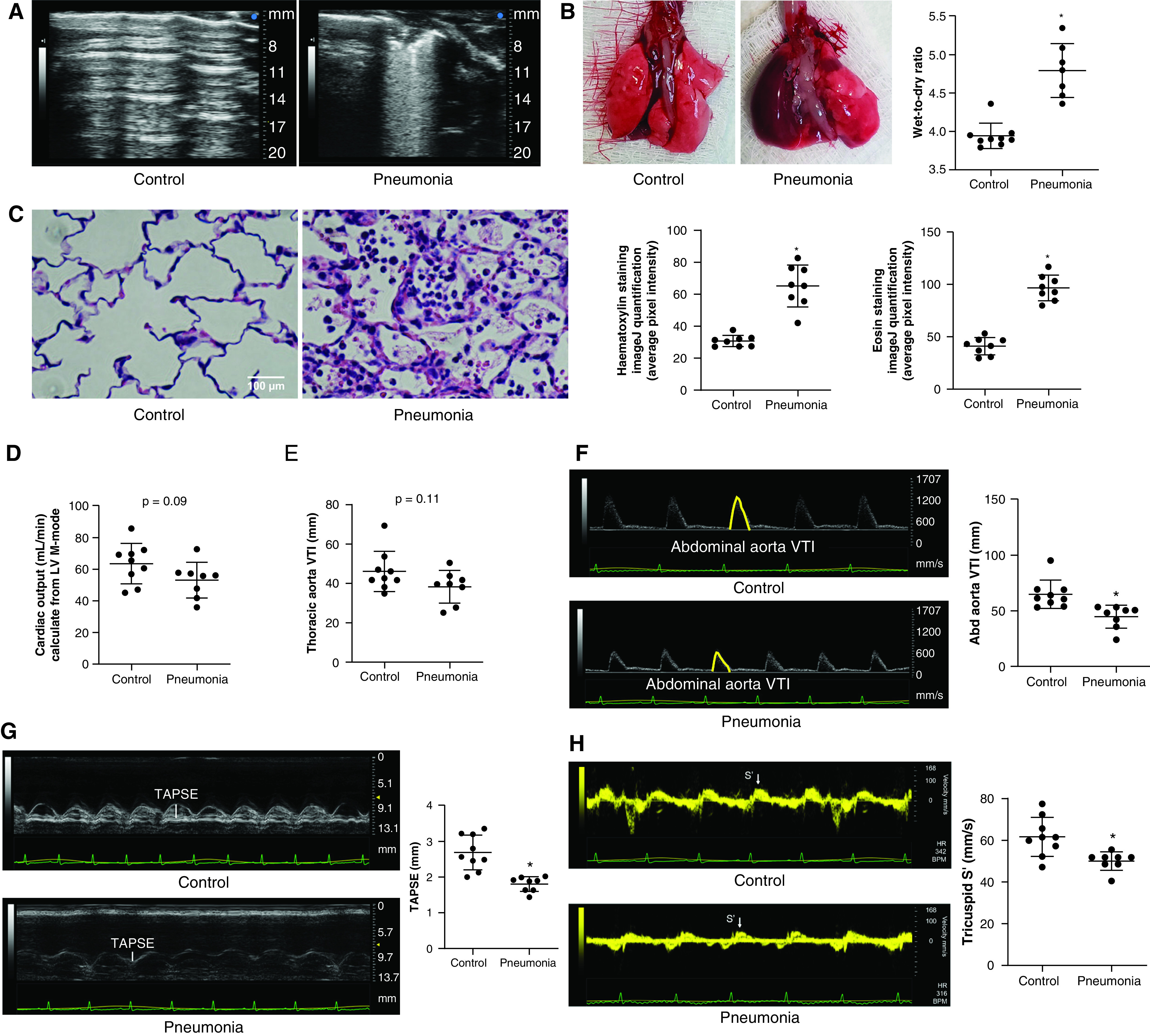
A Pseudomonas aeruginosa pneumonia rat model induces heterogeneous lung injury and cardiovascular changes mimicking human sepsis. To mimic human pneumonia in rats, P. aeruginosa (1 × 10⁷ cfu in 200 μl of PBS) was introduced into the tracheas of Fischer rats. Twenty-four hours later, pneumonia rats (A) developed multiple areas of consolidation and B-line patterns on lung ultrasound images, (B) showed an increased lung tissue wet-to-dry ratio, and (C) showed an increased degree of inflammation as demonstrated by using lung tissue hematoxylin and eosin staining, compared with PBS control rats. For hematoxylin and eosin staining, four evenly spaced axial sections were made throughout the left lungs obtained from two rats per group. Each dot represents an average of two images from each section. Echocardiography revealed no significant change in (D) the cardiac output or(E) the thoracic aortic flow as estimated by using the velocity time integral (VTI), but the (F) Abd aortic VTI, (G) TAPSE, and (H) tricuspid S′ were decreased, indicating potential redistribution of blood flow away from lower body and compromised right ventricular systolic function in pneumonia rats. Scale bar, 100 μm. Data represent the mean ± SD from n = 8–9 animals per group. A Student’s t test was used to compare different groups. *Significant difference (P < 0.05) from control. Abd = abdominal; cfu = colony-forming unit; LV = left ventricular; S′ = tissue Doppler systolic positive wave velocity; TAPSE = tricuspid annular plane systolic excursion.
