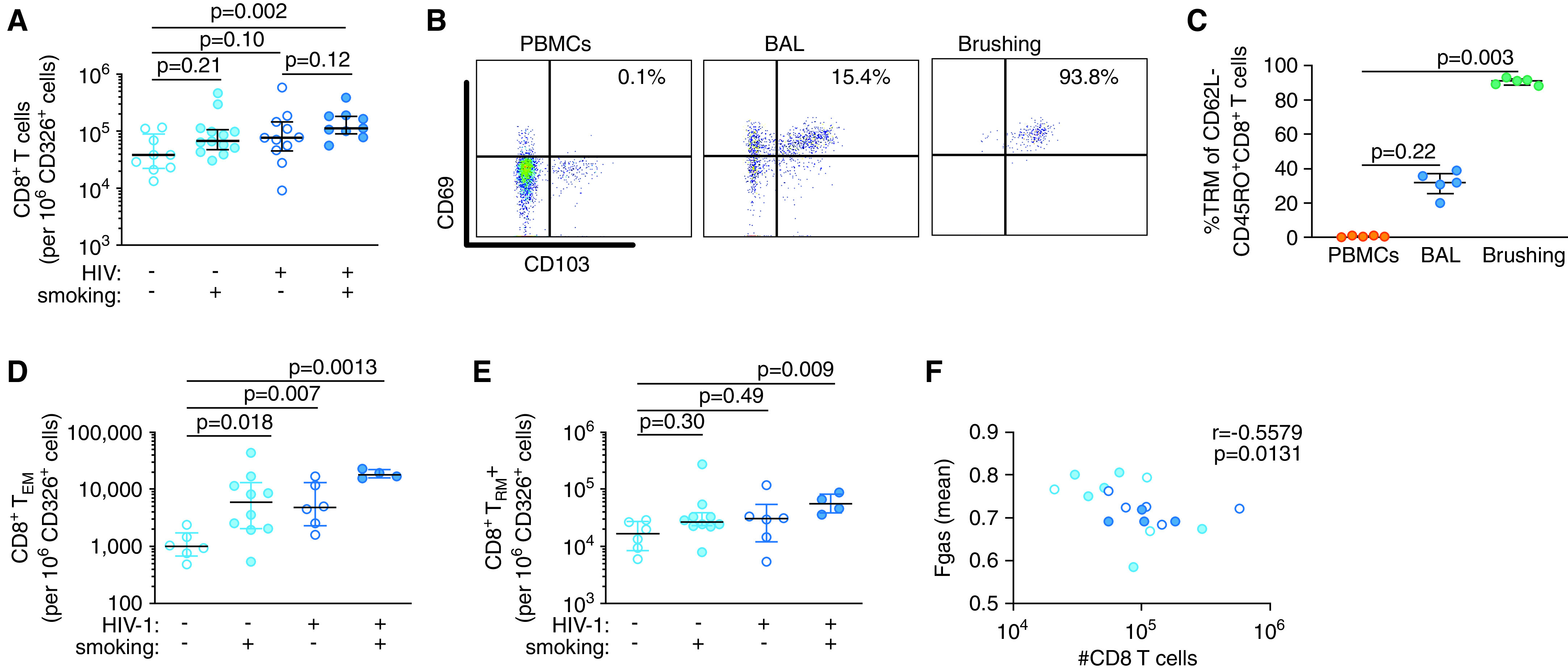
Figure 5.

CD8+ T cells are increased in the airway mucosa of HIV-1–infected smokers. (A) The number of mucosal CD8+ T cells per 106 CD326+ epithelial cells in endobronchial brush samples. (B) Representative flow cytometry plots of CD8+ tissue-resident memory T (TRM) cells in blood, BALF, and endobronchial brush samples. (C) Frequency of CD8+ TRM cells in blood, BALF, and endobronchial brush samples. Number of CD8+ (D) TEM cells and (E) TRM cells in endobronchial brush samples per 106 CD326+ epithelial cells. (F) Correlation of mucosal CD8+ T-cell numbers and mean Fgas as a measure of lung aeration. P values were determined by using nonparametric one-way ANOVA with the Dunn’s test for multiple comparisons (in A and C–E) and the Spearman’s correlation test (in F). Scatter plots are labeled with the median and interquartile range. Fgas = fraction of gas content; PBMC = peripheral blood mononuclear cell; TEM = effector memory T cells.
