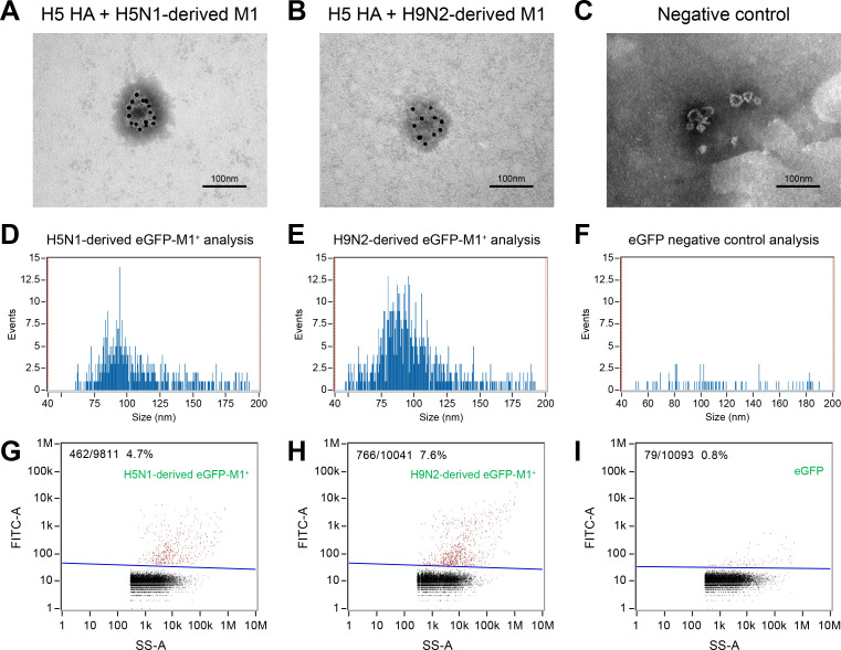
Fig 4. H9N2 virus-derived M1 protein increased viral progeny particle release in mammalian cells.
(A–C) IEM analysis of influenza VLPs in the concentrated cell culture supernatant. 293T cells were co-transfected with (A) HA and H5N1-derived M1, (B) HA and H9N2-derived M1, or (C) empty vector as negative control. Anti-H5N1 IAV HA antibody was used as the primary antibody. The gold is 10 nm in diameter. (D–I) Size and molecular profiling of eGFP-M1+ VLP particles via a Flow NanoAnalyzer. 293T cells were co-transfected with (D, G) HA and H5N1-derived eGFP-M1, (E, H) HA and H9N2-derived eGFP-M1, or (F, I) empty vector as negative control. eGFP was used to label the VLPs. Size profiling of the purified eGFP-M1+ VLP particles with the FITC channel, where Y-axis represents the events, and X-axis represents the size. (G–I) Molecular profiling of eGFP-M1+ VLP particles. SS-A, side scatter area.

