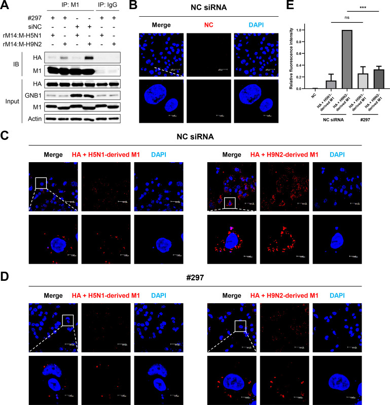
Fig 8. GNB1 facilitated interaction between M1 and HA proteins in mammalian cells.
(A) A549 cells were transfected with NC siRNA or #297 siRNA and infected with recombinant H5N6 virus (rM14:M-H5N1 or rM14:M-H9N2). At 24 hpi, cell lysates were immunoprecipitated using an anti-influenza M1 antibody and probed with an anti-influenza HA antibody. Cell lysates of infected A549 cells were immunoprecipitated using an anti-IgG antibody and probed with an anti-influenza HA antibody as negative control. Compared to the normal cells, the interaction between H9N2-derived M1 and HA proteins was decreased in the GNB1-silenced cells. IB, immunoblot. (B–D) Influenza M1 and HA proteins interaction was determined by PLA. The PLA was performed using antibodies specific to influenza M1 and HA proteins. The fluorescence of cells was analyzed by a fluorescence confocal microscope (red fluorescent signal). Nuclei were stained with DAPI (blue). (B) PLA of the cells transfected with NC siRNA and an empty vector. (C) PLA of the cells transfected with NC siRNA, HA, and H9N2- or H5N1-derived M1 encoding plasmids. (D) PLA of the cells transfected with #297 siRNA, HA, and H9N2- or H5N1-derived M1 encoding plasmids. (E) Multiple images (B–D) were processed by BlobFinder to measure the PLA fluorescence intensity per cell (~30 cells total for each condition). The graphs show the means ± SD of three independent experiments normalized to the HA+H9N2-derived M1 (NC siRNA) PLA signal. (***, P < 0.001; ns, no significance.)

