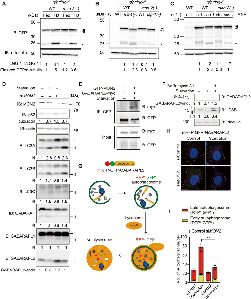
Fig. 6. MON-2/MON2 appears to increase autophagy by activating LGG-1/GABARAPL2 in C. elegans and mammalian cells.
(A to C) mon-2(xh22) [mon-2(−)] consistently decreased the high level of cleaved GFP caused by food deprivation (FD) (A), cco-1 RNAi (B), and isp-1(qm150) [isp-1(−)] mutations (C) (n = 3). Different from consistent results for cleaved GFP, LGG-1–II/I ratio was somewhat variable likely because of subtle separation of LGG-1–I and II bands. Filled arrowhead, LGG-1–I. Open arrowhead, LGG-1–II. Asterisk, cleaved GFP. α-Tubulin, loading control. WT without a transgene, negative control. IB, immunoblot. (D) Small interfering RNA (siRNA) targeting MON2 (siMON2) substantially increased the level of GABARAPL2 while having small effects on those of other tested autophagosomal markers (p62, LC3A, LC3B, LC3C, GABARAP, and GABARAP L1) under basal or starvation conditions (n = 2). One endogenous GABARAPL2 band was detected, which was decreased under starvation and increased by siMON2. In contrast, phosphatidylethanolamine-conjugated (II) and nonconjugated proteins (I) were separated for the other ATG8/LC3 family members. (E) GABARAPL2 was coimmunoprecipitated with MON2 under starvation conditions (n = 3). IP, immunoprecipitation. (F) Immunoblot for GABARAPL2, LC3B, and vinculin in HeLa cells after starvation or starvation with bafilomycin A1 treatment for 6 hours (n = 2). (G) Illustrated fluorescence changes in mRFP-GFP-GABARAPL2 based on its subcellular location in early or late autophagosomes, or autolysosomes. (H) siMON2 in HeLa cells stably transfected with mRFP-GFP-GABARAPL2 under basal or starvation conditions altered the flux of GABARAPL2. Scale bars, 10 μm. (I) The numbers of RFP+GFP+ puncta and RFP+GFP− puncta were counted for each cell and analyzed for statistical differences (N = 21, 21, 22, and 16). Error bars represent SEMs **P < 0.01, Mann-Whitney test).

