
Figure 4. In Situ Assay on Demethylation Activity of KDM5C.
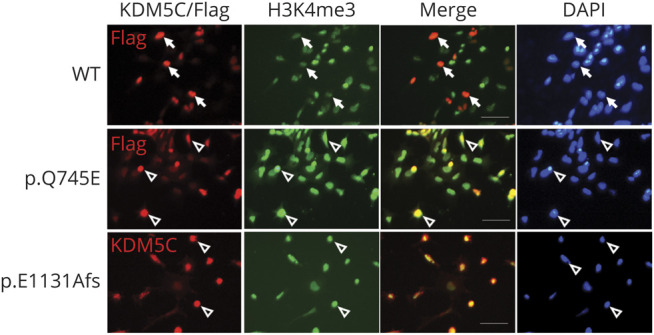
Representative immunofluorescence images of KDM5C and H3K4me3 in cells transfected with WT and variations of KDM5C (N = 4). Cells transfected with WT KDM5C showed reduced H3K4me3 signals, implicating demethylase activity of KDM5C (arrow). Cells transfected with p.Q745E or p.E1131Afs variation retained high H3K4me3 signals implicating the reduction of demethylase activity of KDM5C (open arrowhead). Nuclei were stained with DAPI. Scale bar = 50 μm. H3K4 = H3 lysine 4