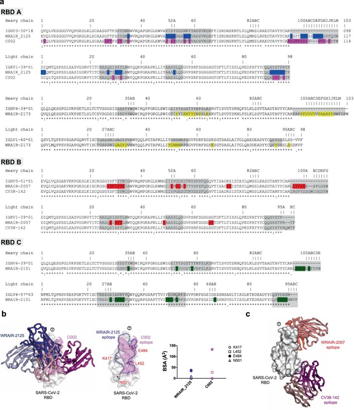
Extended Data Fig. 6. Structure and sequence alignment.
a, Sequence alignment of RBD A, B and C WRAIR mAbs with their precursor germline genes. Antibody residue numbers are labelled on the top and CDRs are shaded gray. All antibody residue numbering and CDR loops are designated using the Kabat numbering system. Antibody residues that interact with RBD are colored as antibody structures in Fig. 2. Symbols *,:, and. denote identical, similar and less similar residues, respectively. Previously published antibodies, C002 and CV38-142, with identical germline-encoded genes as WRAIR-2125 and WRAIR-2057, respectively, are also aligned. b, Left panel: C002 structure (magenta) is overlaid onto the WRAIR-2125 (dark blue) structure. Middle panel: Frequently occurring SARS-CoV-2 VOC residues are shown as sticks on the RBD surface with WRAIR-2125 and C002 epitopes highlighted in dark blue and magenta colors, respectively. Right panel: Buried surface area (BSA) for VOC residues, related to the mAbs WRAIR-2125 and C002 are shown as dot plots. T symbol is used to designate the “tip” of the RBD molecule. c, MAb CV38-142 structure (purple) is overlaid onto the WRAIR-2057-RBD complex structure (red).

