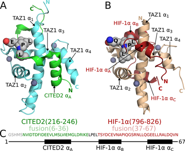
Figure 1. TAZ1:CITED2 and TAZ1:HIF-1α structures and the design of a CITED2-HIF-1α fusion.
(A) NMR structure of the TAZ1:CITED2 complex (PDB 1R8U; (De Guzman et al., 2004)). TAZ1(CBP 340–439) is shown in cyan and CITED2(220–269) is shown in green. Zinc atoms are shown as dark-gray spheres. Side chains of the CITED2 LPEL motif are shown as light-gray spheres with N and O atoms colored blue and red respectively. Black labels are superimposed on the LPEL side chains. TAZ1 and CITED2 helices and N- and C-termini are labeled for reference. Residues 262–269 of CITED2 are omitted for clarity. (B) NMR structure of the TAZ1:HIF-1α complex (PDB 1L8C; (Dames et al., 2002)). TAZ1(CBP 345–439) is shown in tan and HIF1α(776–826) is shown in red. Zinc atoms are shown as dark-gray spheres. Side chains of the HIF-1α LPQL motif are shown as light-gray spheres with N and O atoms colored blue and red respectively. Black labels are superimposed on the LPQL side chains. TAZ1 and HIF-1α helices and N- and C-termini are labeled for reference. (C) Amino acid sequence of the fusion peptide. Sequences from CITED2 (residues 216–246) and HIF-1α (residues 796–826) are colored green and red respectively and correspond to residues 6–36 and 37–67 of the fusion peptide. The LPEL motif of the CITED2 sequence is shown in black. The N-terminal GSHMS sequence is a cloning artifact (see methods). The block diagram below the sequence denotes the positions of the CITED2 and HIF-1α helical binding motifs.

