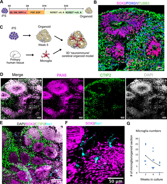
Figure 2. ‘Neuroimmune’ Organoid Model of Human Brain Development.
A. Cortical organoids were generated and pattern using dual SMAD and Wnt inhibition. After day 35 in culture, cortical organoids were moved into a maintenance media containing vitamin A and were maintained in the same media for the duration of the experiment. B. Cortical organoids express forebrain marker FOXG1, radial glia marker Sox2 and pan-neuronal marker TUBB3. C. Organoids were generated from iPSC cells and after switching to maintenance media at five weeks were combined with primary human microglia to generate neuro-immune organoids. D. Cortical organoids express markers for cortical radial glial PAX6 and cortical layer 5 neuronal marker CTIP2. E. Microglia distribute within the organoid. F. Iba1-positive microglia cells are ramified and distributed between and around rosettes. G. Microglia numbers monotonically decrease over the five-week co-culture time.
See also Figure S4.

