
Figure 4. Microglia transplanted into organoids recapitulate primary human microglia.
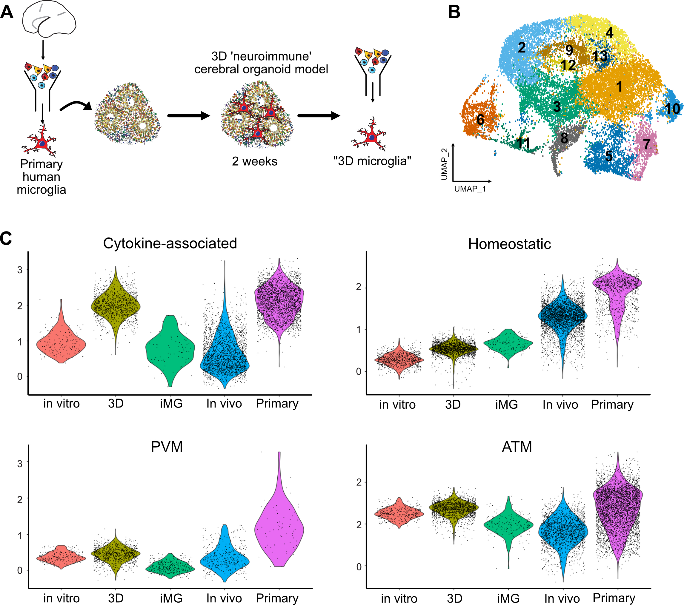
A. Primary human microglia from primary human GW23 brain were purified using MACS Cd11b beads and added to cortical organoids. Neuro-immune organoids were allowed to develop for two weeks, and then microglia cells were re-purified using CD11b magnetic beads for downstream scRNAseq processing. B. 17,705 microglia cells purified from organoids formed 13 clusters. C. Cross-model comparison of primary human microglia cultured in vitro (“in vitro”), primary human microglia engrafted in cortical organoids (“3D”), induced microglia (“iMG”), induced microglia transplanted into mouse brain (“In vivo”), and primary human microglia from mid-gestation from the BICCN dataset.
See also Figure S5.