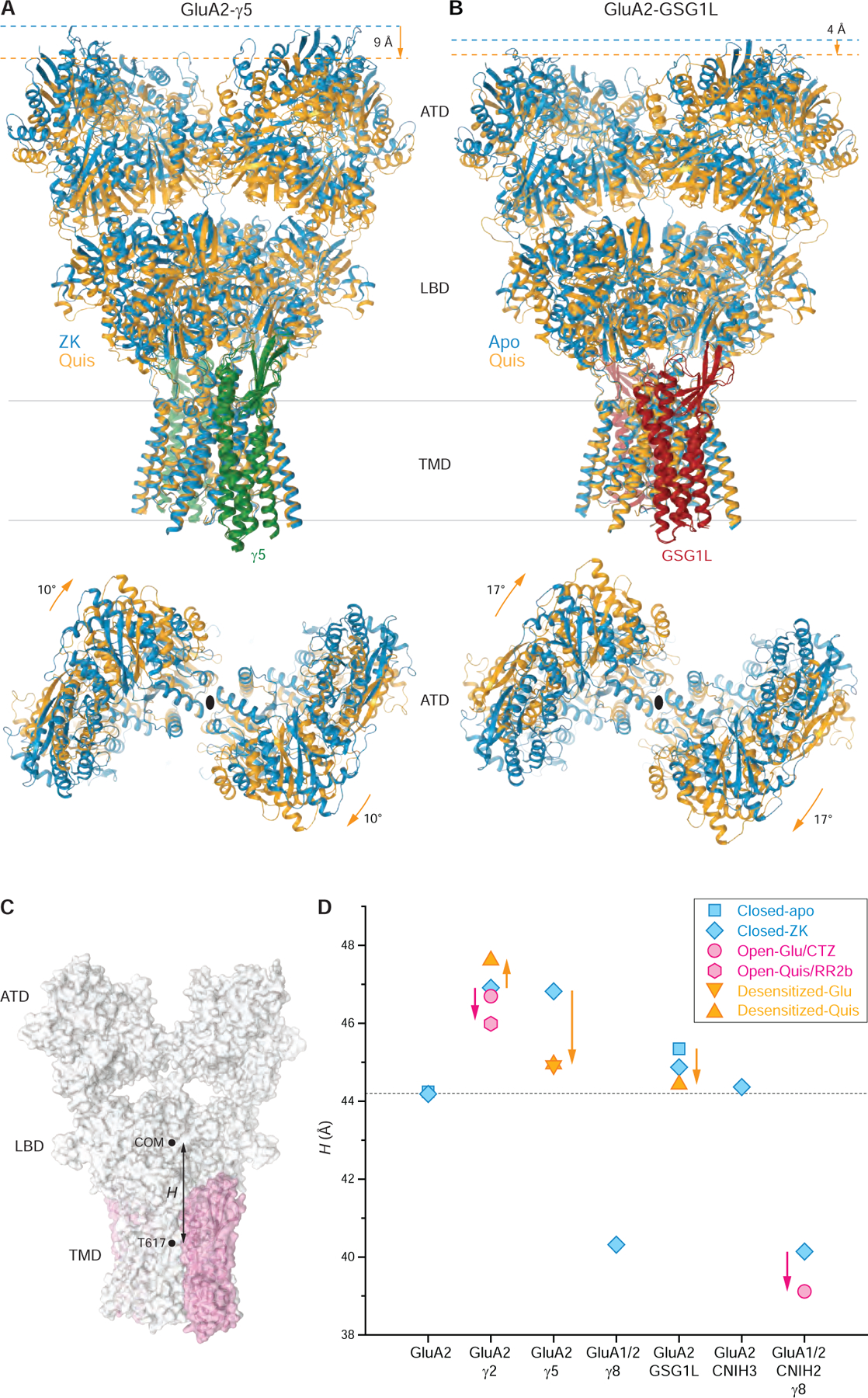
Figure 6. Different closed to desensitized state transformations of GluA2-γ5 and GluA2-GSG1L complexes and elevation of LBD over TMD in the presence of different auxiliary subunits.

(A-B) TMD-based superposition of GluA2-γ5 (A) and GluA2-GSG1L (B) in the closed (blue) and desensitized (orange) states viewed parallel to the membrane (top) or extracellularly (bottom), with γ5 shown in green and GSG1L in red. Shortening of the receptor and ATD rotation that signify the desensitized to closed state conversion are indicated by orange arrows. Note, in the closed state, GluA2-γ5 is ~4 Å taller than GluA2-GSG1L.
(C) Schematic illustrating the measurement of LBD over TMD elevation (H) as a distance between the center of mass (COM) for the LBD layer and the T617 Cα coordinates averaged over four AMPA receptor subunits.
(D) LBD over TMD elevation for GluA2 alone and AMPA receptor complexes with different auxiliary subunits in the closed (blue), desensitized (orange) and open (pink) states. Measurements are for the same structures as in Figure 5G, including the structures reported in this study, GluA2-γ5ZK, GluA2-GSG1Lapo, GluA2-γ5Glu, GluA2-γ5Quis, and GluA2-GSG1LQuis, as well as the previously published structures GluA2apo (PDB ID: 5L1B), GluA2ZK (PDB ID: 3KG2), GluA2-γ2ZK (PDB ID: 5KK2), GluA2-γ8ZK (PDB ID: 6QKC), GluA2-CNIH3ZK (PDB ID: 6PEQ), GluA2-GSG1LZK (PDB ID: 5WEK), GluA2-γ2Quis (PDB ID: 5VOV), GluA2-γ2Glu+CTZ (PDB ID: 5WEO), GluA1-GluA2-γ8-CNIH2ZK (PDB ID: 7OCE), GluA1-GluA2-γ8-CNIH2Glu+CTZ (PDB ID: 7OCF) and GluA2-γ2Quis-RR2b (PDB ID: 5VOT). Changes in H accompanying transitions from the closed to desensitized and open states are indicated by orange and pink arrows, respectively.
See also Figure S7.
