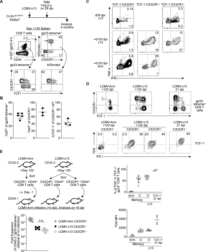
FIGURE 6. CX3CR1+ CD8 T cells during the chronic phase give rise to TCF-1+ chronic memory cells following the resolution of viremia.
(A-B) Flow cytometry plots showing expression of tdTomato (tdT), TCF-1, CX3CR1 and TIM3 in gp33-specific CD8 T cells in the splenocytes of LCMV-c13-infected Cx3cr1creERT2/+ R26tdT mice treated with Tamoxifen on 28 dpi and analyzed on >120 dpi. Representative data from two independent experiments with n=3 mice is shown. Flow plots are shown in (A) and replicates from one experiment are shown as mean ± SD. (B). (C) Expression of IFN-γ and TNF-α in indicated subsets of CD8 T cells in the spleen of 29 dpi LCMV-c13-infected mice, >120 dpi LCMV-Armstrong-infected mice, or >120 dpi LCMV-c13-infected mice following stimulation with LCMV-gp33 peptide ex vivo for four hours. Data are representative of two independent experiments with n=XX mice each (D) Expression of TCF-1 and CX3CR1 in LCMV-gp33-specific splenic CD8 T cells (top) and expression of PD-1 and TOX in TCF-1+ LCMV-gp33-specific CD8 T cells at indicated time point after LCMV-Arm or LCMV-c13 infection. Frequencies of TOX+ cells and levels of TOX expression are shown with mean ± SD. (E) Schematic of adoptive transfer experiment to assess the capability of secondary expansion of CX3CR1+ and CX3CR1− CD8 T cells that were sorted from LCMV Arm and LCMV-c13 infected mice. The right panel shows data from replicates. Data are representative from 2 independent experiments with n=2 donor mice and n=5 recipient mice per experiment. Fold expansion was calculated by fold change of gp33-specific CD8 T cells followed by normalization to no-infection control.

