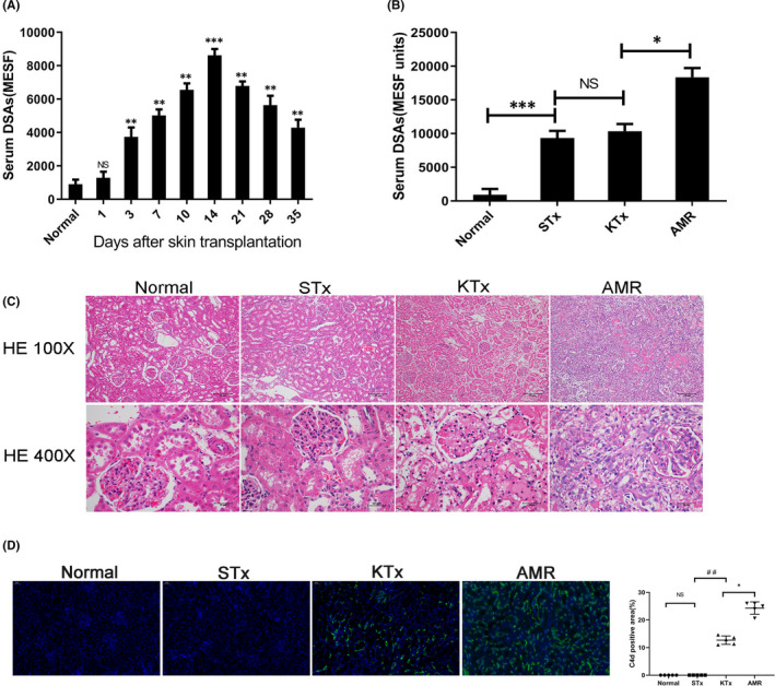
FIGURE 1.

Constructing and identifying a rat model of AMR following kidney transplantation. Brown Norway (BN) rats were pre‐sensitized with Lewis (LEW) rats by skin transplantation to construct an antibody‐mediated rejection (AMR) model for kidney transplantation. Kidney grafts were harvested on day 4 after kidney transplantation. NS, no significance; *p < 0.05; ** p < 0.01; *** p < 0.001; ## p < 0.01. (A) Serum IgG level of BN rats after skin transplantation; mean fluorescence intensity (MFI) was converted to molecules of equivalent soluble fluorochrome (MESF) units. The level of donor‐specific antibodies (DSAs) reached peak level on day 14 after skin transplantation. (B) Serum IgG level of BN rats in the normal group, STx (skin transplantation) group, KTx (kidney transplantation) group and the AMR group. (C) Histopathology of the kidneys. Compared with the normal group, the STx group and the KTx group, the kidneys in the AMR group showed significant peritubular capillaries (PTC) inflammation and glomerulitis. (D) C4d staining of the kidneys. Compared with the normal group, STx group and the KTx group, C4d staining on PTCs was positive on day 4 in the AMR group following transplantation
