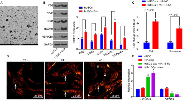
Figure 4.

miR‐16‐5p is released to HPDCs via hUSC exosomes and effectively inhibits VEGFA in HPDCs. A, TEM was used to observe the morphology change in hUSCs‐Exo (scale bar = 200 nm). B, the expression of CD9, CD63, CD81, TSG101 and HSP90B1 was determined by Western blot analysis. C, RT‐qPCR was used to determine miR‐16‐5p expression in hUSCs and hUSC exosomes. D, the uptake of hUSC exosomes by HPDCs was observed at different time‐points (12 h, 24 h, and 48 h), white arrows pointed to the CFSE‐labelled exosomes (green), while HPDCs were red‐stained (scale bar = 25 μm). E, expression of miR‐16‐5p and VEGFA was determined in each group by RT‐qPCR. *P < .05 vs the HPDC group. Measurement data were expressed as mean ± standard deviation. Independent sample t test was used for comparison between two groups, and multivariate analysis of variance was employed in the comparison between multiple groups. The experiment was repeated three times. CFSE, carboxyfluorescein diacetate succinimidyl ester; Exo‐depl, hUSCs co‐cultured with HPDCs in a exosome‐depleted medium; HPDCs, human podocytes; hUSCs, human urine‐derived stem cells; miR‐16‐5p, microRNA‐16‐5p; RT‐qPCR, reverse transcription‐quantitative polymerase chain reaction; TEM, transmission electron microscopy; VEGFA, vascular endothelial growth factor A
