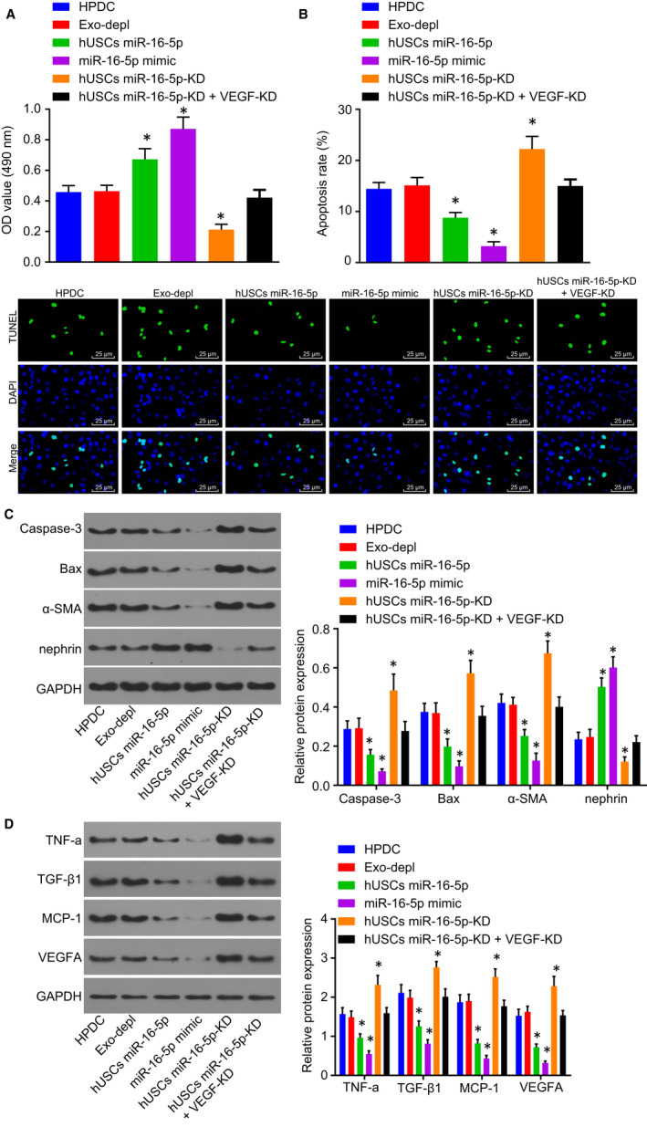
Figure 5.

Secretion of miR‐16‐5p from hUSC exosomes ameliorates podocyte injury induced by HG. A, the viability of podocytes was detected by CCK‐8 assay. B, TUNEL was performed to measure the apoptosis rate of podocytes (scale bar = 25 μm). C, Western blot analysis was applied to determine the expression of nephrin, α‐SMA, Bax and Caspase‐3. D, the expression of VEGFA and relate factors MCP‐1, TGF‐β1 and TNF‐α examined by Western blot analysis. *P < .05 vs the HPDC group. Measurement data were expressed as mean ± standard deviation. Multivariate analysis of variance was used for comparison among multiple groups, and the experiment was repeated 3 times. CCK‐8, Cell Counting Kit‐8; Exo‐depl, hUSCs co‐cultured with HPDCs in a exosome‐depleted medium; HG, high glucose; HPDCs, human podocytes; hUSCs miR‐16‐5p‐KD + VEGF‐KD, hUSCs transfected with miR‐16‐5p inhibitor and siRNA‐VEGFA plasmid and co‐cultured with HPDCs; hUSCs miR‐16‐5p‐KD, hUSCs transfected with miR‐16‐5p inhibitor and co‐cultured with HPDCs; hUSCs, human urine‐derived stem cells; MCP‐1, monocyte chemoattractant protein‐1; miR‐16‐5p, microRNA‐16‐5p; TGF‐β1, transforming growth factor‐β1; TNF‐α, tumour necrosis factor‐α; TUNEL, Terminal deoxynucleotidyl transferase (TdT)‐mediated 2′‐Deoxyuridine 5′‐Triphosphate (dUTP) nick‐end labelling; VEGFA, vascular endothelial growth factor A
