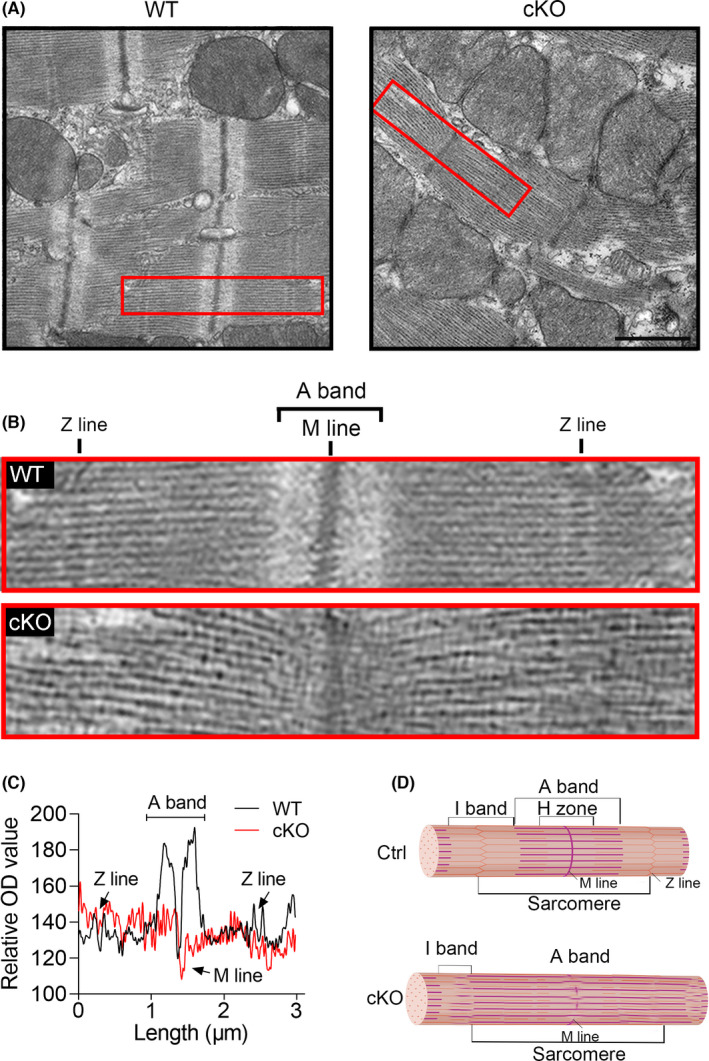
FIGURE 6.

YTHDC1 deficiency destroyed the ultrastructure of the cardiac sarcomere. (A)–(B) Representative transmission electron microscopy (TEM) of sarcomere structure (top, scale bar: 1μm) from Ctrl and cKO mice at 8‐week‐old. The red box indicates an intact sarcomere. (C) The optical density (OD) analysis for the single sarcomere showing that Ythdc1 deficiency destroyed the normal ultrastructure of the cardiac sarcomere. (D) The schematic representation of a cardiac sarcomere. The lateral boundaries of the sarcomere are the Z‐discs. The I‐bands surrounds the Z‐disc and is a region where thin filaments are not superimposed by thick filaments. The A‐band region contains thin filaments and thick filaments. The M‐band falls within the H‐zone, where thick filaments do not interdigitate with thick filaments
