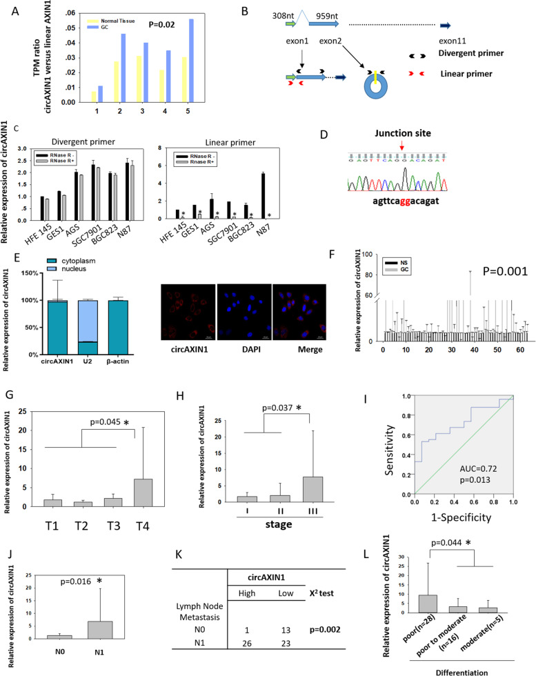
Fig. 3.
Characterization of circAXIN1. a The TPM ratio of circAXIN1 versus linear AXIN1 is elevated in GC samples obtained from a cohort of patients in the ATGC database. b An illustration of the linear AXIN1 and circAXIN1 genomic regions. CircAXIN1 is formed from the head to tail splicing of exon2 of the AXIN1 parental gene. The specific divergent and convergent primers were designed as shown to detect circAXIN1 and linear AXIN1, respectively. c CircAXIN1 is expressed at a higher level in GC cells compared with that in normal gastric epithelial cells. CircAXIN1 is resistant to RNase R-digestion but linear AXIN1 is not. d Sanger sequencing was performed to validate the back splicing of circAXIN1 using divergent primers. e CircAXIN1 is mostly localized in the cytoplasm. Left: cytoplasmic and nuclear RNA was extracted, and quantitative PCR was performed to detect the relative subcellular localization of circAXIN1. U2 was used as the positive control for nuclear localization and β-actin for cytoplasmic localization. Right: Using probes specific to the junction site, FISH indicated that circAXIN1 is localized in the cytoplasm. f CircAXIN1 is highly expressed in GC tissues (p = 0.001). NS: Normal Sample. g CircAXIN1 is highly expressed in T4 compared with its expression in T1–3. h CircAXIN1 is overexpressed in stage III compared with its expression in stage I and II. i Receiver operating characteristic (ROC) curve analysis of normalized circAXIN1 expression of GC with and without lymph node metastasis. The area under the ROC curve (AUC) conveys the accuracy, in terms of its sensitivity and specificity, of this biomarker in distinguishing GC lymph node metastasis. j CircAXIN1 is significantly highly expressed in GC with lymph node metastasis. k The expression of circAXIN1 is positively associated with lymph node metastasis. l CircAXIN1 is particularly highly expressed in poorly differentiated tumors compared to moderately differentiated and moderately to poorly differentiated tumors with lymph node metastasis

