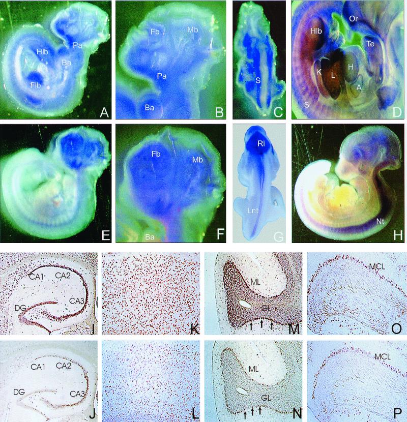
FIG. 9.
In situ hybridization of whole-mount mouse embryos and adult mouse brain sections. (A to H) Expression patterns of profilin I (A to D) and profilin II (E to H) during early embryonic development. (A, B, E, and F) Lateral view of E10.5 embryos. Profilin I (A and B) is found in the midbrain (Mb), forebrain (Fb), branchial arches (Ba), pharyngeal arches (Pa), forelimb bud (Flb), and hind limb bud (Hlb). Profilin II expression (E and F) was detected in the entire brain region and the neural tube. (C and G) Dorsal view of E10.5 embryos shows profilin I expression in the somites (S) and profilin II in the rhombic lip (Rl) and the lateral neural tube (Lnt). (D and H) Lateral views of E11.5 embryos showing profilin I expression (D) in the olfactory region (Or), tongue epithelium (Te), heart (H), atrium (A), liver (L), kidney (K), somites, and hind limb bud and profilin II expression (H) in the developing brain and the neural tube (Nt). (I to P) Overlapping expression of profilin I and profilin II transcripts in adult mouse brain. Coronal (I to L) or sagittal (M to P) paraffin sections were hybridized with digoxigenin-labeled antisense riboprobes specific for profilin I (I, K, M, and O) or profilin II (J, L, N, and P). (I and J) Profilins I and II are expressed in the dentate gyrus (DG) and the CA regions of the hippocampus. (K and L) Strong expression of profilins was detected in all layers of the cortex. (M and N) Intensive signals were observed in the Purkinje cells (arrows) of cerebellum. Cells in both the molecular (ML) and granular (GL) layers were positive for profilins I and II. (O and P) Main olfactory bulb; strong staining was seen in the mitral cell layer (MCL).

