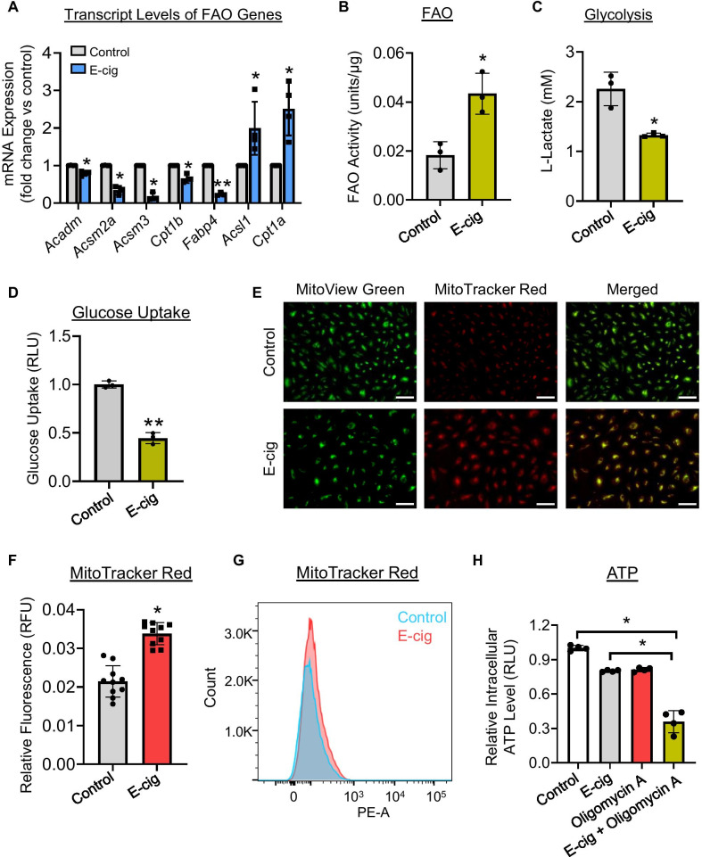
Fig. 6.
Assessment of metabolic changes in iPSC-ECs following e-cig treatment. A qPCR validation of FAO genes following e-cig treatment (6.5 TPE). B FAO, C glycolysis, and D glucose uptakes of iPSC-ECs following e-cig exposure (6.5 TPE) for 24 h (n = 3). Representative images and corresponding quantitative data of control and e-cig-treated cells taken with a fluorescence microscope (E, F) or flow cytometry (G). Live cells were stained with MitoTracker Red and MitoView Green. H ATP levels in cells treated with vehicle or e-cig (6.5 TPE) and/or 1.5 µM of oligomycin A for 48 h. Data are represented as mean ± SD, and * and ** indicate p < 0.05 and p < 0.001, respectively. Scale bars = 100 μm

