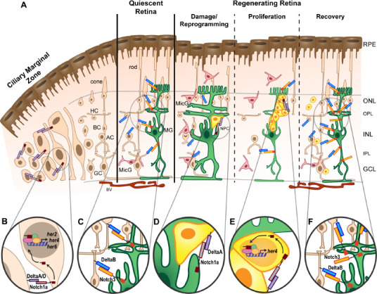
Figure 1.

Dynamic Notch signaling during retinal regeneration.
(A) Schematic of the adult zebrafish retina. The retina is organized into three nuclear layers: ganglion cell layer (GCL), inner nuclear layer (INL), and outer nuclear layer (ONL), which are separated by the inner plexiform layer (IPL) and outer plexiform layer (OPL) where synaptic connections are located. Müller glia (MG) cell bodies reside in the INL, but their projections extend throughout all three nuclear layers. MG are mostly quiescent in the intact retina, except for occasional proliferation to give rise to rod photoreceptor progenitors. In the intact, quiescent retina, resting microglia (MicG, pink) reside in the plexiform layers with ramified morphology. The inner, basal surface of the retina is vascularized (blood vessels, BV), and the outer, apical surface is lined with the retinal pigment epithelium (RPE). The ciliary marginal zone (CMZ) at the periphery of the retina is an area of persistent neurogenesis with proliferative multipotent retinal progenitors that give rise to ganglion cells (GC), amacrine cells (AC), bipolar cells (BC), horizontal cells (HC), and cone photoreceptors. (B) The CMZ is marked by expression of notch1a and notch1b receptor genes, deltaA and deltaD ligand genes, and downstream target genes her2, her4, and her6 (see text). (C) MG express Notch3, which when activated with the DeltaB ligand, maintains MG quiescence. Upon damage, neurons degenerate, and MG hypertrophy, followed by reprogramming and asymmetric cell division to retain a MG and generate a neuronal progenitor cell (NPC). MicG migrate to areas of damage and adopt an amoeboid morphology. (D) Notch3 expression is downregulated in the proliferating retina, but other Notch signaling components are necessary for continued proliferation of the NPCs, in particular Notch1a and DeltaA. (E) Continued NPC proliferation produces a cluster of NPCs in the regenerating retina. This process requires the Notch downstream target, her4. (F) The NPCs migrate to the areas of damage and differentiate into the lost neurons. Notch3 expression is reestablished in the quiescent MG.
