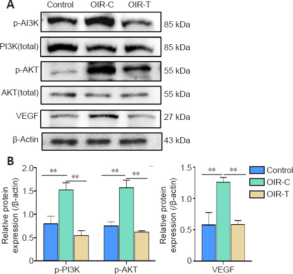
Figure 6.

Protein expression levels of p-PI3K, p-AKT and VEGF in the retina of mice at the age of 17 days.
(A) Western blot bands of PI3K, AKT, p-PI3K, p-AKT, and VEGF proteins. (B) Quantification of p-PI3K, p-AKT, and VEGF proteins. Data are expressed as the mean ± SD (n = 15 mice per group). **P < 0.01 (Mann-Whitney U test). AKT: Serine/threonine kinase; OIR-C: oxygen induced retinopathy, control eye; OIR-T: oxygen induced retinopathy eye injected intravitreally with MEG3 overexpression lentivirus PI3K: phosphoinositide 3-kinase; VEGF: vascular endothelial growth factor.
