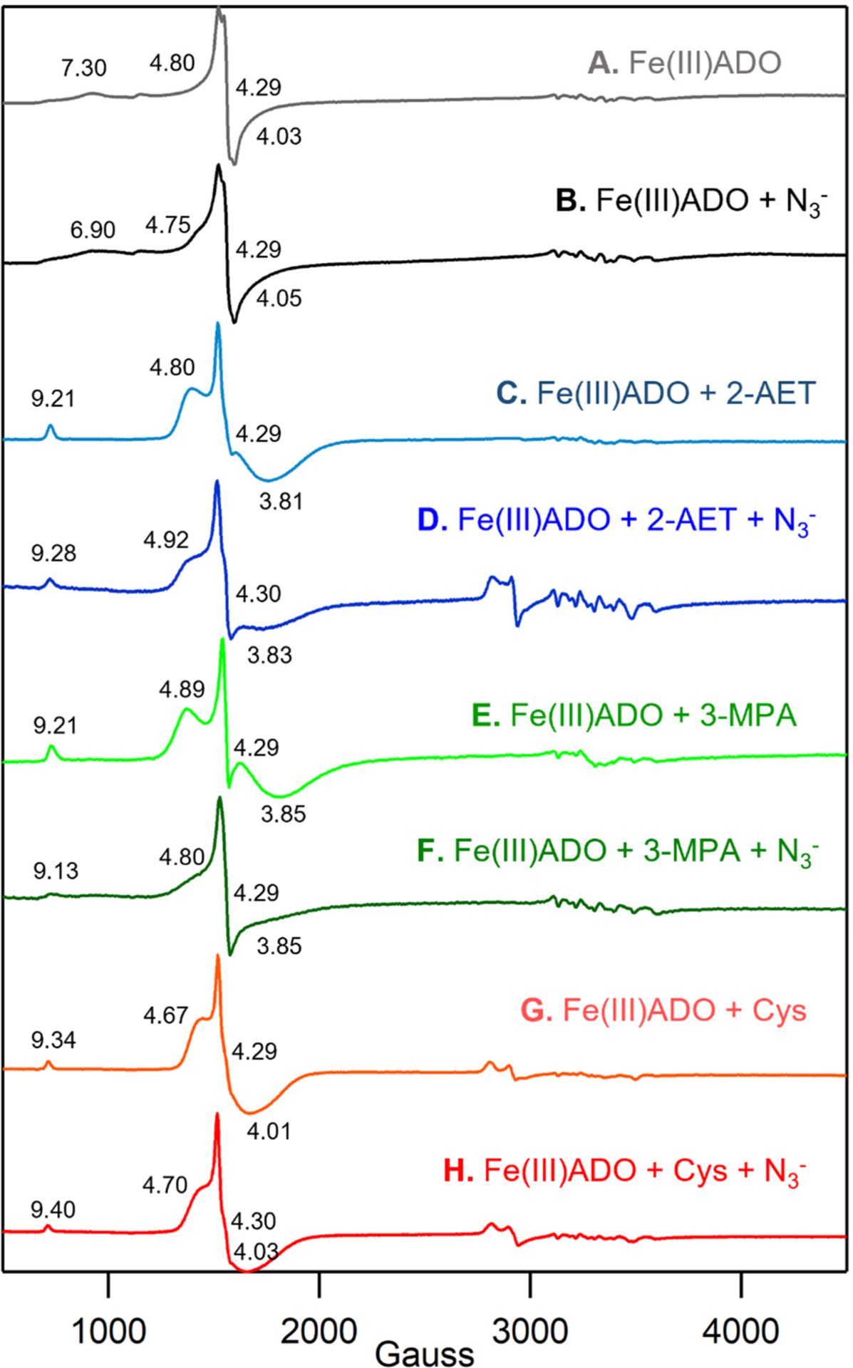
Figure 2.

X-band EPR spectra at 20 K of 0.4 mM Fe(III)ADO in the absence and presence of various substrate analogues and/or O2•− surrogates. A) Fe(III)ADO, B) Fe(III)ADO incubated with a 200-fold excess (80 mM) of azide, C) Fe(III)ADO incubated with a 5-fold excess (2 mM) of 2-AET, D) Fe(III)ADO incubated with a 5-fold excess (2 mM) of 2-AET and a 200-fold excess (80 mM) of azide, E) Fe(III)ADO incubated with a 10-fold excess (4 mM) of 3-MPA, F) Fe(III)ADO incubated with a 10-fold excess (4 mM) of 3-MPA and a 200-fold excess (80 mM) of azide, G) Fe(III)ADO incubated with a 10-fold excess (4 mM) of Cys, and H) Fe(III)ADO incubated with a 5-fold excess (2 mM) of Cys and a 200-fold excess (80 mM) of azide. Effective g values are provided for the S = 5/2 species. The hyperfine structure in the 3000–3700 cm−1 Gauss region is due to a minor Mn(II) impurity.
