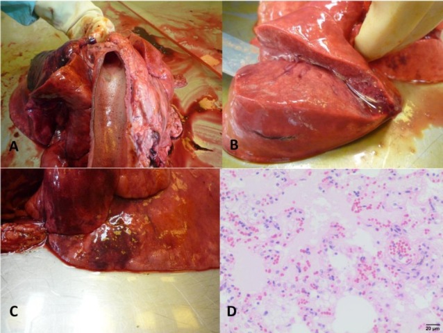
Fig. 1.

Pulmonary changes in dogs infected with B. canis
A – Foamy exudate in the trachea of dog No. II; B – Pulmonary oedema in dog No. II; C – Pulmonary congestion and foci of emphysema in dog No. IV; D – Microphotograph of pulmonary oedema in dog No. VI, visible oedema fluid filling alveoli and dilatation of blood vessels (haematoxylin and eosin staining)
