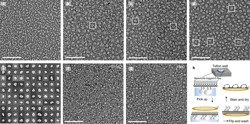
Fig. 5 |. Electron microscopy images with negative stain of K-Ras assembly.
a–d, K-Ras wild type at a 10 μM concentration; maleimide lipids were used at the following percentages: 0% (a), 5% (b), 10% (c) and 20% (d). e, 2D class average of the K-Ras wild-type particles. f, D154Q K-Ras at a 10 μM concentration with 20% maleimide lipids. g, K88D K-Ras at a 10 μM concentration with 20% maleimide lipids. h, Workflow of the experiment. After incubation with the monolayer lipid overnight, the K-Ras particles were moved to a carbon-coated copper grid, washed intensively, negatively stained and examined by electron microscopy. Scale bars, 200 nm.

