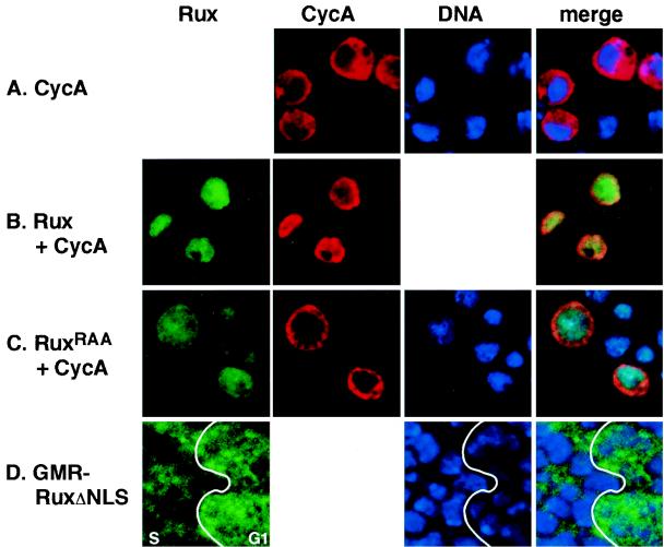
FIG. 1.
Nuclear localization of Rux requires the C-terminal bipartite NLS. Confocal optical sections of Drosophila SL2 cells (A to C) and eye imaginal tissue (D) ectopically expressing CycA (A to C), wild-type Rux (B), and Rux NLS mutant proteins (C and D). CycA expression is shown in red, Rux expression is shown in green, and DNA is shown in blue. (A) CycA is cytoplasmically localized in SL2 cells. (B) Wild-type Rux is localized to the nucleus in SL2 cells. Coexpression of Rux and CycA results in the translocation of CycA to the nucleus. (C) When the Rux NLS is mutated (RuxRAA), CycA remains cytoplasmic while Rux is distributed uniformly throughout the cell. In this example, the first RKR cluster in the Rux NLS has been changed to RAA. (D) Expression of the RuxΔNLS mutant protein in the eye imaginal disk. The junction between the G1 domain in the MF (G1) and the synchronous domain of S-phase cells behind the MF (S) is demarcated by a line. G1 cells show both nuclear and cytoplasmic expression of RuxΔNLS, while the mutant protein is down-regulated in the nuclei of cells reentering S phase. The anterior edge of the disk is to the right.

