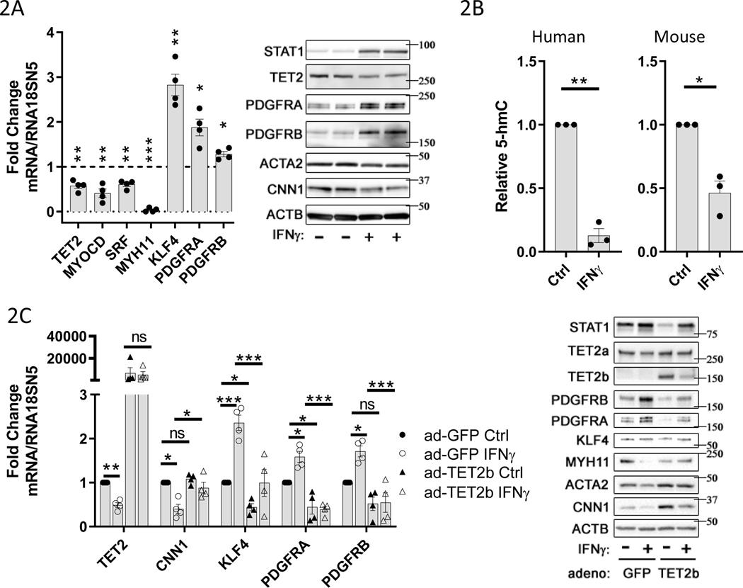
Figure 2: IFNγ treatment causes TET2 and 5-hmC repression, and an activated phenotype in VSMCs.
Primary human or mouse vascular smooth muscle cells (VSMCs) were cultured under control conditions (Ctrl or “-“) or treated with 100ng/ml recombinant human or mouse IFNγ (IFNγ or “+”) for 48hrs, respectively. A) Left panel: mRNA levels for indicated genes from hVSMCs determined by quantitative PCR (qPCR) normalized to RNA18SN5, expressed as fold change relative to control (dotted line) (N=4), Right panel: representative western blot (N=4). B) Quantitation of genomic 5-hmC content, by ELISA, 48hr after treatment of VSMCs with 100ng/ml IFNγ. Left panel: human VSMCs (N=3), Right panel: mouse VSMCs (N=3). C) hVSMCs were infected with adenoviruses expressing GFP control or TET2b protein prior to IFNγ treatment as described in 2A-B. Left panel: mRNA levels for indicated genes determined by qPCR normalized to RNA18SN5 (N=4), Right panel: representative western blot (N=4). A-B) one sample t test, C) Two-way ANOVA: *P<0.05, **P< 0.01, ***P<0.001.

