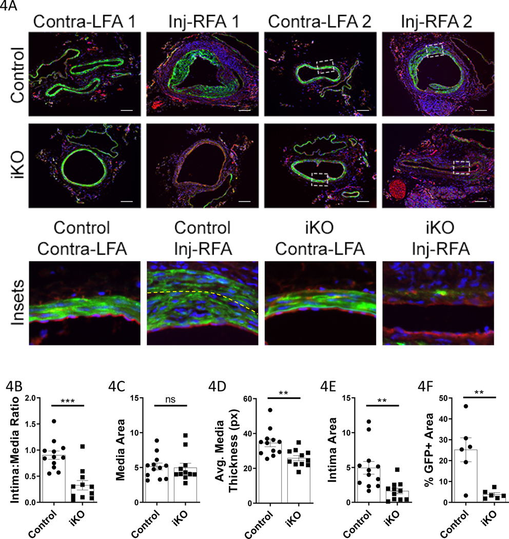
Figure 4: TET2-deficient femoral arteries exhibit medial thinning, VSMC loss, and limited intimal hyperplasia in response to endothelial denudation injury.
Guidewire-injured right femoral arteries (Inj-RFA), and contralateral control left femoral arteries (Contra-LFA), of littermate Tet2++Myh11-Cre (Control N=6; Control mTmG-labeled N=6) and Tet2FFMyh11-Cre (iKO N=5; iKO mTmG-labeled N=6) were harvested 21days after surgery. A) Representative micrographs of uninjured (LFA) and injured (Inj-RFA) vessels. Scale bar= 100μm. Below: high power insets (indicated by white dashed line in lower power images). Yellow dashed line indicates the internal elastic lamina in the control Inj-RFA sample. B) Quantification of intima:media ratio from both mTmG-labeled and unlabeled animals (Control N=12, iKO N=11). C) Quantification of media area from both mTmG-labeled and unlabeled animals (Control N=12, iKO N=11). D) Quantification of average media layer thickness from both mTmG-labeled and unlabeled animals (Control N=12, iKO N=11). Medial thickness was measured at eight points (0°, 45°, 90°, 135°, 180°, etc.) around the circumference of each vessel and averaged. E) Quantification of intima area from both mTmG-labeled and unlabeled animals (Control N=12, iKO N=11). F) Quantification of %GFP+ area in the media and intima from mTmG-labeled animals only (Control N=6, iKO N=6). B-D Student’s t test, E-F Welch’s t test: **P<0.01, ***P<0.001.

