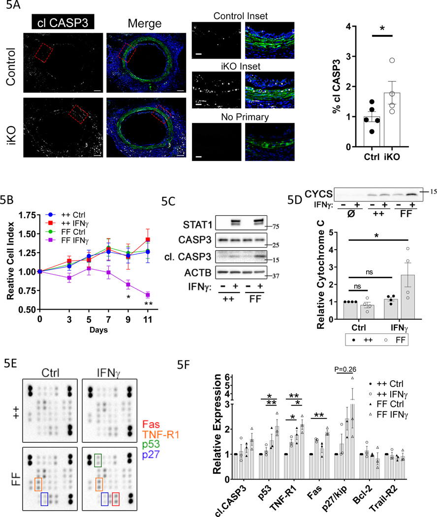
Figure 5: Depletion of Tet2 increases VSMC apoptosis in response to GA and IFNγ through upregulation of mediators of extrinsic apoptosis.
Control and TET2-deficient thoracic aorta segments from male donor mice were transplanted into sex-mismatched recipients of the same strain. A) Micrographs of GFP fluorescence and cleaved caspase 3 (clCASP3) immunostaining in littermate Tet2++mTmGmut+Myh11-Cre (Ctrl; N=5) and Tet2FFmTmGmut+Myh11-Cre (iKO; N=4) grafts 10 days after surgery; Right) quantitation of cl.CASP3-positive area within GFP-positive cells (% clCASP3) in the media-intima combined region. Red bars indicate the medial layer. B-F) mVSMCs isolated from Tet2++mTmGmut+Myh11-Cre (++) and Tet2FFmTmGmut+Myh11-Cre (FF) mice were treated with Cre recombinase-encoding adenovirus in vitro. ++ and FF cells were then cultured under control conditions (Ctrl or “-“) or treated with 100ng/ml rm-IFNγ (IFNγ or “+”) at day 0. B) Viability was assessed by crystal violet staining from 0– 11 days after IFNγ treatment (N=6). Western blot of VSMC C) whole-cell lysates or D) conditioned medium treated from mVSMCs treated with IFNу for 7days as in A. Ø indicates medium conditioned for 7 days in the absence of cells. In C), total STAT1 upregulation (long or short exposure) serves as a positive control known response to IFNγ treatment. E) Apoptosis antibody array of VSMC lysates treated with IFNу for 5 days. F) Quantitation of apoptosis array (N=3). A) Student’s t test: *P<0.05. B, D,F) Two-way ANOVA *P<0.05, **P<0.01.

