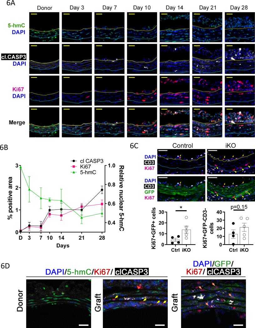
Figure 6: TET activity is negatively correlated with apoptosis and proliferation in GA.
Thoracic aorta segments from male donor mice were transplanted into sex-mismatched recipients of the same strain. A) Micrographs of immunostained sections from donor (N=5) and graft vessels at 3 (N=3),7 (N=3),10 (N=3),14 (N=2),21 (N=4), and 28days (N=4) after surgery. Upper panels) Ki67 and cleaved caspase 3 (clCASP3), and lower panels) 5-hmC. Representative images shown. B) Quantification of Ki67 and clCASP3 % positive area (left y-axis) from micrographs as described in A. Quantification of nuclear 5-hmC signal (right y-axis) from the same tissues as described in A. C) 10day Control and Tet2-deficient graft sections were immunostained for Ki67, CD3, and GFP. Left graph) Ki67 positive (Ki67+) donor derived VSMCs (GFP+) and right graph) Ki67 positive non-donor derived VSMC (GFP–), non-T cells (CD3–) were quantified in the media-intima combined region. Student’s t test: *P<0.05. D) Donor and graft sections were immunostained for 5-hmC or GFP, Ki67, cleaved caspase 3 (clCASP3), and DAPI. Yellow arrows indicate 5-hmClo Ki67+ cells, orange arrows indicate GFP+Ki67+ cells. Representative images of 4 independent samples are shown.

