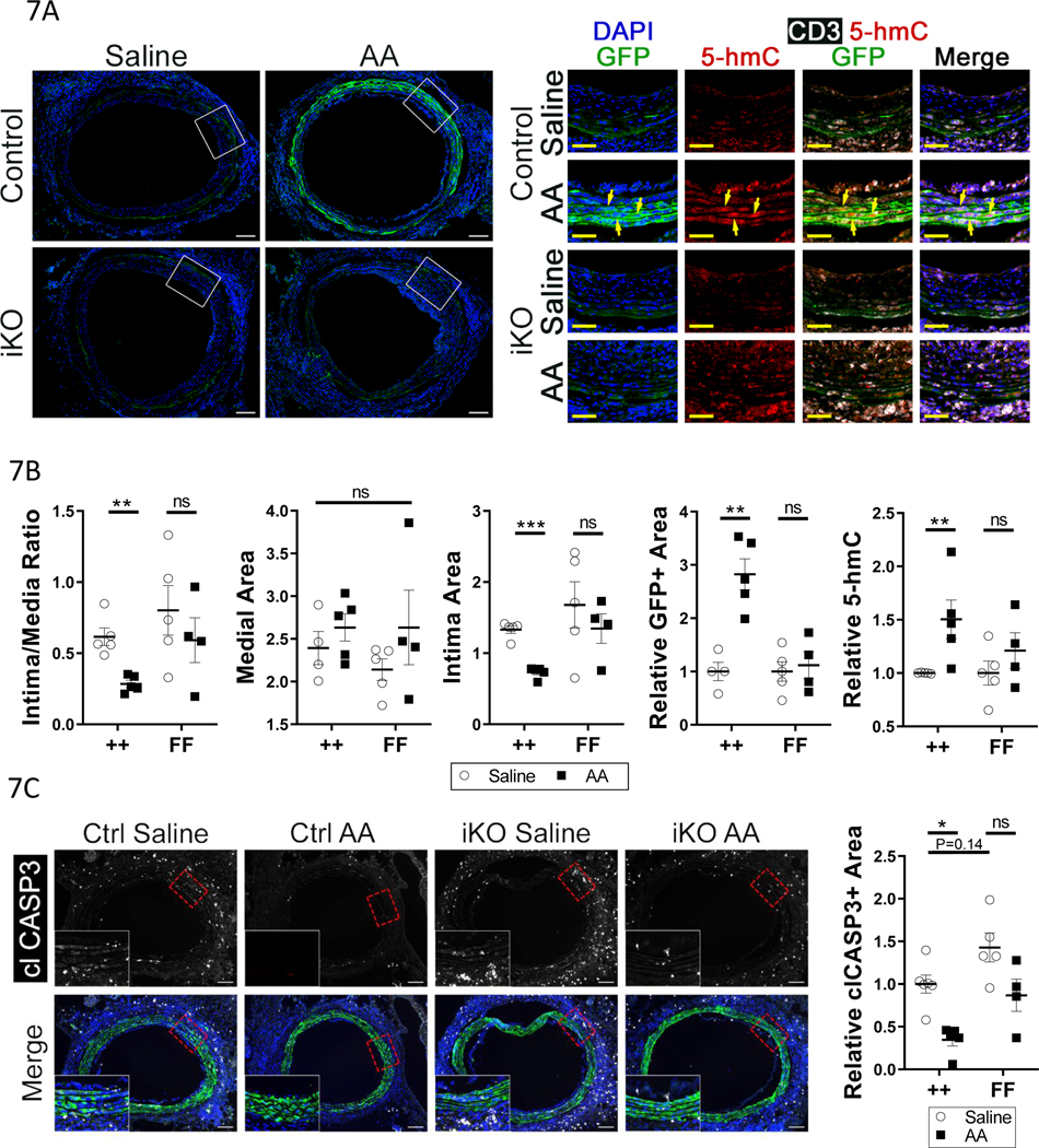
Figure 7: Promoting TET2 activity with ascorbic acid rescues GA-induced donor-VSMC apoptosis and decreases intimal hyperplasia.
A) Micrographs of GFP fluorescence and DAPI, 28 days after surgery, in littermate Tet2++mTmGmut+Myh11-Cre (Control) and Tet2FFmTmGmut+Myh11-Cre (iKO) grafts in C57bl/6J recipients receiving either saline or 4mg/kg/day ascorbic acid (AA). Right) High-power insets of areas in panel A denoted by dashed boxes; micrographs of 5-hmC, CD3, and GFP immunostaining. Yellow arrows indicate GFP+ VSMCs with high nuclear 5-hmC. B) Quantification of intima:media ratio, media area, intima area, relative GFP positive (GFP+) area in the media/intima, and relative nuclear 5-hmC intensity in the media/intima, in grafts from control and TET2-deficient animals treated with saline or AA. C) Left) Micrographs of graft tissues -performed as in 7A-B but for 10 days – were immunostained for cleaved caspase 3 (clCASP3). Right) quantification of clCASP3+ area in GFP+ cells, relative to ++ saline. A-B) ++ saline N=5, ++ AA N=5, FF saline N=5, FF AA N=4. C) ++ saline N=6, ++ AA N=5, FF saline N=5, FF AA N=4. Two-way ANOVA: *P<0.05, **P< 0.01, ***P<0.001.

