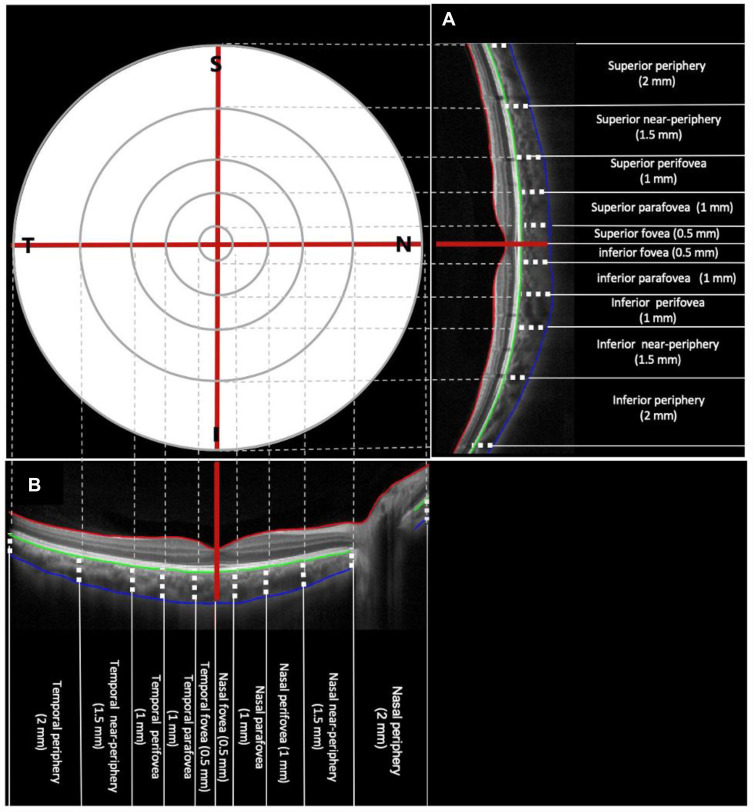
Figure 1.
Vertical (A) and horizontal (B) single line image obtained with optical coherence tomography (OCT) instrument, the foveal pit as manually marked in the custom-written software (straight red line in (A and B), the anterior (green line in (A and B) and posterior (blue line in (A and B) are the boundaries of the choroid.

