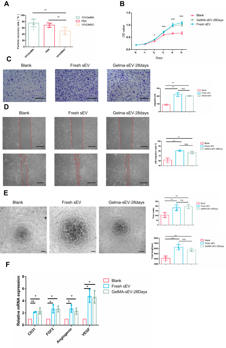
Figure 4.
The effect of preserved sEV on HUVEC proliferation, migration, tube formation and angiogenesis differentiation. (A) Recovery rate of sEV from different preservation method by TEI isolation. (B) Proliferation of HUVEC co-cultured with fresh sEV and sEV preserved in GelMA hydrogels for 28 days. n = 3 for each group. (C) Images of migrated HUVECs in different group. Scale bar = 200μm. n = 3 for each group. (D) Images of scratch assay in different group. Scale bar = 100μm. Time transition of the percentage of cell-free zone against initial scratch area after 12 hour, n = 3 for each group. (E) Tube-like structures of HUVECs in different group. Scale bar = 200 µm. n = 3 for each group. (F) The expression of angiogenesis markers (VEGF, FGF2, CD31, Angiogenin) was detected by qRT-PCR at 4 days post sEV treatment, n = 3 for each group. The significance (A–E) was tested with one-way ANOVA with Tukey posthoc test. (*p < 0.05, **p < 0.01, ***p < 0.001).

