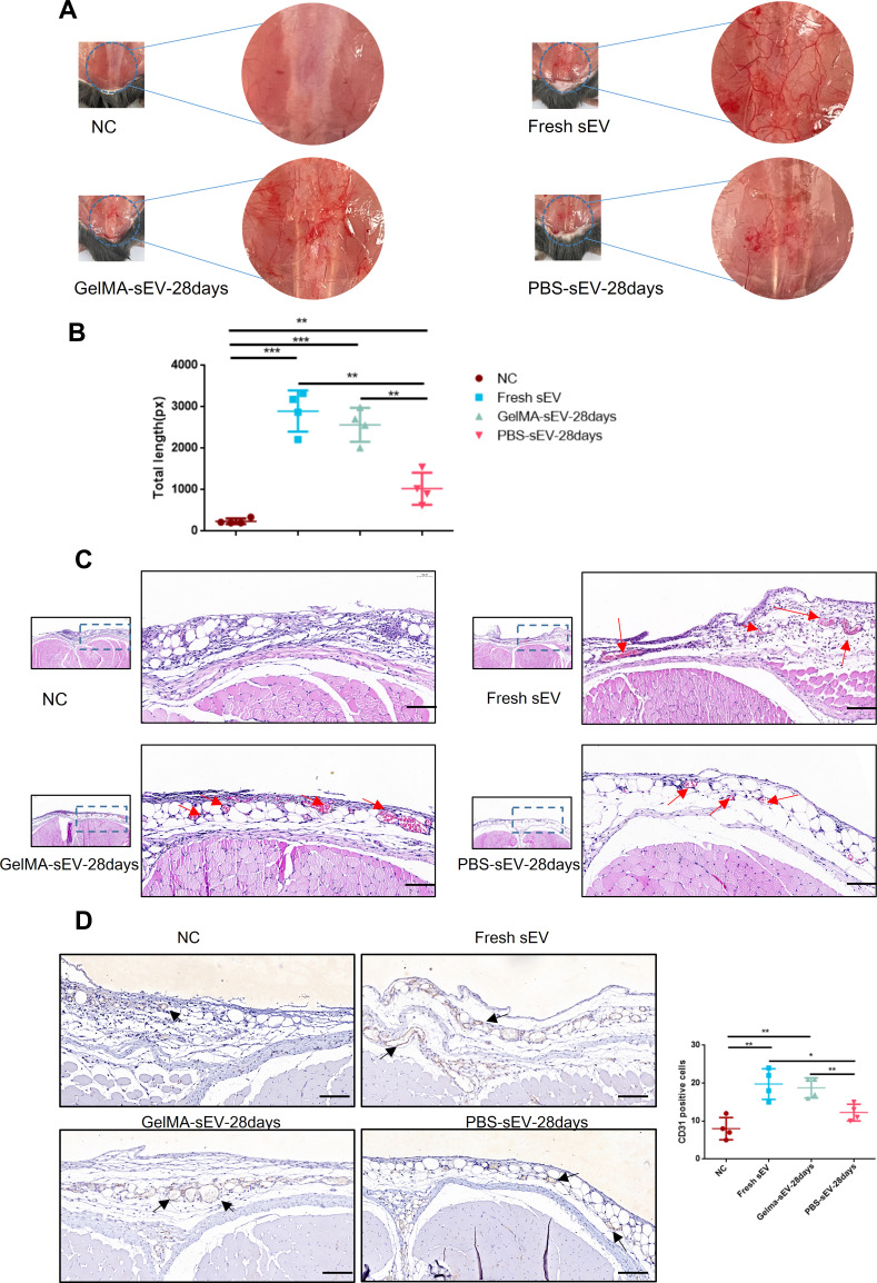
Figure 6.
Preserved GelMA-sEV complex promoted angiogenesis in the subcutaneous fascial layer of C57BL/6J mice. (A) General view of angiogenesis in the subcutaneous fascial layer in different groups on 6th day after treatment.NC, negative control. (B) Total length of new vessels in the fascial layer from each groups, n = 4. (C) Representative HE-stained images of angiogenesis in the subcutaneous fascial layer (red arrows: new vessels with erythrocytes) Scale bar = 50µm. (D) Immunohistochemical staining of CD31-positive endothelial cells in each groups was used to visualize vessels. CD31-positive endothelial cells were quantified from 5 independent visions per sample (black arrows: CD31 positive endothelial cells) Scale bar = 50 µm, n = 4. The significance (A and D) was tested with one-way ANOVA with Tukey posthoc test. (*p < 0.05, **p < 0.01, ***p < 0.001).

