
Figure 4. Physiological Role of NLRP6 Phase Separation in Anti-RNA Virus Defense in Hepatocytes.
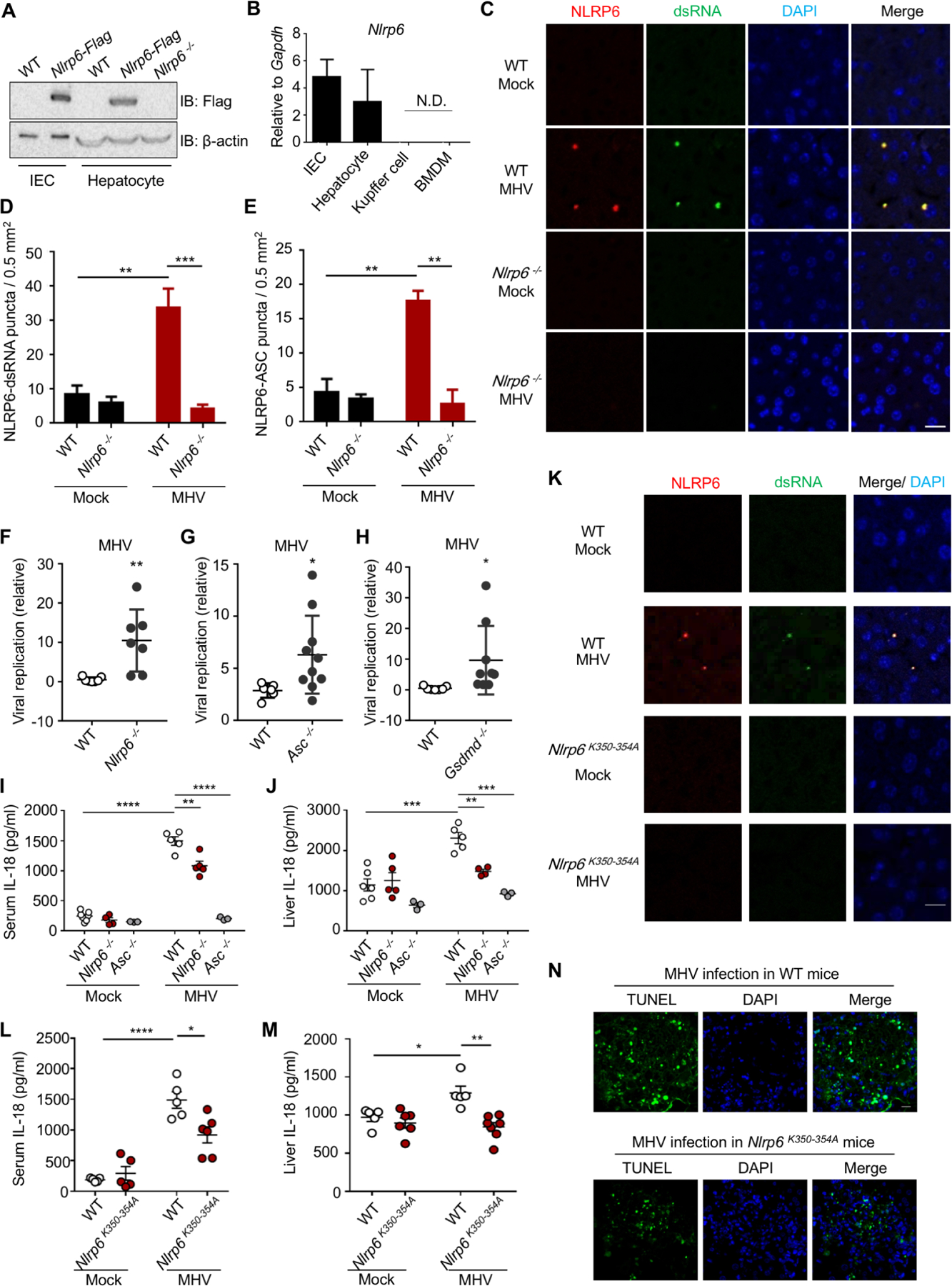
WT, Nlrp6−/− or Nlrp6K350−354A knock-in mice were infected with MHV (i.p.) and analyzed as indicated (C–N).
(A) Immunoblot of NLRP6 in primary hepatocytes or intestinal epithelial cells (IECs) from Nlrp6-Flag knock-in mice.
(B) qPCR assays for Nlrp6 mRNA levels in different cells. N.D.: not detectable.
(C) Fluorescence immunohistochemistry staining of dsRNA (J2) and NLRP6 in liver sections from uninfected or MHV-infected WT and Nlrp6−/− mice. Nuclei were stained with DAPI.
(D, E) Quantification of NLRP6-dsRNA (D) and NLRP6-ASC (E) puncta in liver sections. Data are mean ± SEM from 6 randomly sampled regions.
(F-H) Viral replication in the liver 3 days after MHV infection in Nlrp6−/− (F), Asc−/− (G) and Gsdmd−/− mice (H) in comparison to WT mice.
(I, J) Serum (I) and liver-explant supernatant (J) IL-18 levels determined by ELISA from WT, Nlrp6−/− and Asc−/− mice 3 days post-infection.
(K) Fluorescence immunohistochemistry staining of dsRNA (J2) and NLRP6 in liver sections from uninfected or MHV-infected WT and Nlrp6K350−354A mice. Nuclei were stained with DAPI.
(L, M) Serum (L) and liver-explant supernatant (M) IL-18 levels in WT and Nlrp6K350−354A mice 3 days post-infection.
(N) TUNEL staining of MHV-infected WT and Nlrp6K350−354A mice 3 days after infection. Scale bar: 20 μm.