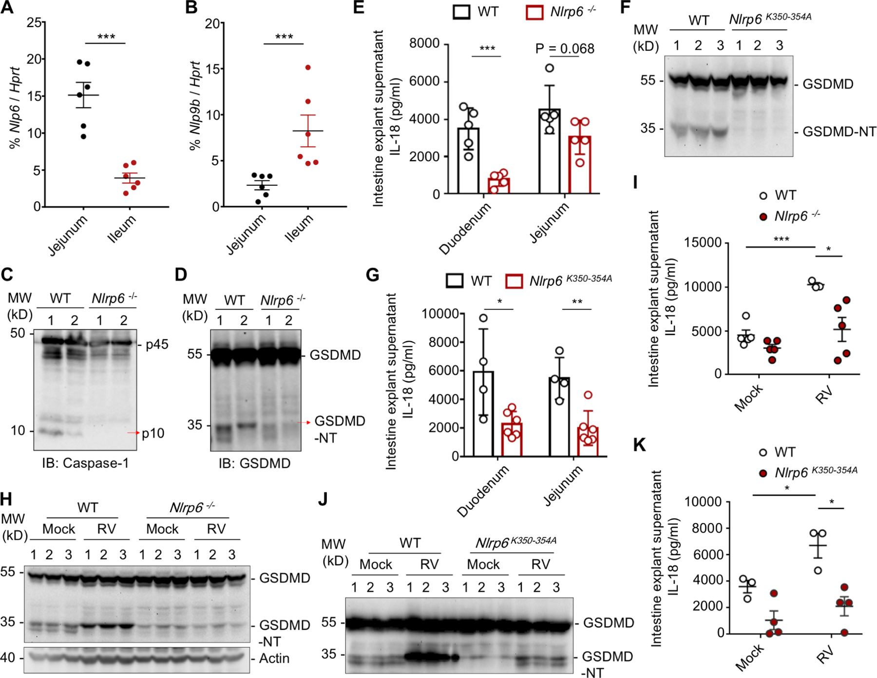
Figure 5. The Physiological Role of NLRP6 Phase Separation in IECs in Vivo.

(A, B) qPCR assays for Nlrp6 (A) and Nlrp9b (B) mRNA levels in different locations of the intestine relative to the Hprt control. Mean ± SEM for 6 mice is displayed for each group.
(C, D) Immunoblotting of IEC lysates of multiple WT and Nlrp6−/− mice for caspase-1 (C) and GSDMD (D).
(E) IL-18 levels of intestine explant supernatant of WT and Nlrp6−/− mice determined by ELISA. Mean ± SEM for 5 mice is displayed for each group.
(F) Immunoblotting of IEC lysates of 3 WT and K350–354A mutant mice each for GSDMD processing.
(G) IL-18 levels of intestine explant supernatant of WT and K350–354A mutant mice determined by ELISA. Mean ± SEM for 4 WT and 6 mutant mice is displayed for each group.
(H) Immunoblot of 3 small intestine homogenates each from mock or RV-infected WT and Nlrp6−/− mice 2 days post RV infection to detect cleaved GSDMD.
(I) Intestine-explant supernatant IL-18 levels in WT and Nlrp6−/− mice 2 days post-infection.
(J) Immunoblot of 3 small intestine homogenates each from mock or RV-infected WT and K350–354A mutant mice 2 days post RV infection to detect cleaved GSDMD.
(K) Intestine-explant supernatant IL-18 levels in WT and K350–354A mutant mice 2 days post-infection.
