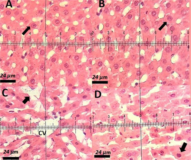
Fig. 9.

A Hematoxylin-eosin-stained section of a control rat showing characteristic normal liver histopathology. The arrow indicates a normal hepatocyte with a normal nucleus and cytoplasm. H&E staining (×40); B Liver sections of rat vacuolation (arrow).H&E staining (×40); C Liver section of a PTX-treated rat, a decrease in cytoplasmic vacuolization, arrow point indicates the hepatocytes regeneration. H&E staining (×40); D Liver section of a rat treated with niosomal PTX, a decrease in pyknotic nuclei, H&E staining (×40)
