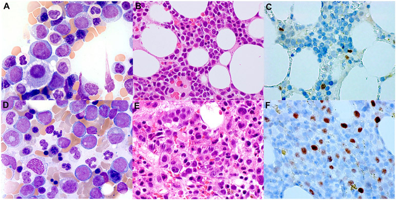
Figure 1. MYC immunohistochemistry (IHC) staining in MDS and AML-MRC patients.
The representative images of low MYC expression in the bone marrow interpreted with MDS-EB-I (A-C) and high MYC expression in the bone marrow diagnosed with AML-MRC (D-F). Wright-Giemsa (A and D, 1000x), H&E (B and E, 600x), and MYC staining (C and F, immunoperoxidase, 600x).

