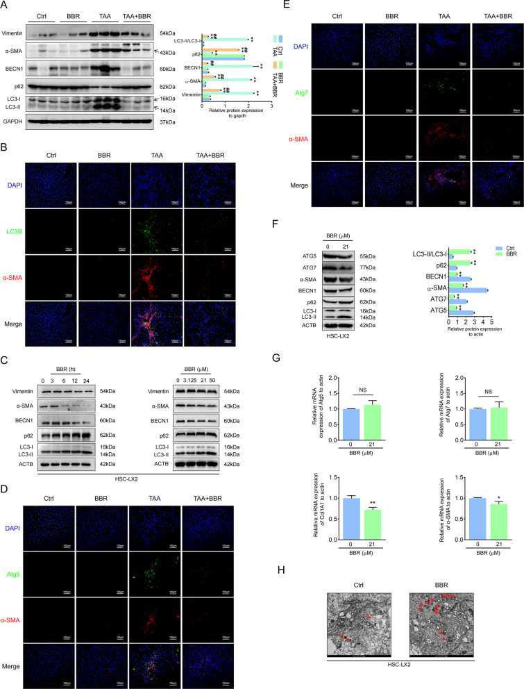
Fig. 4. Autophagy is involved in BBR-induced depression of HSC activation in liver fibrosis.
A The expression levels of BECN1, p62, LC3B, vimentin, and α-SMA were detected by western blot in TAA-induced mouse liver fibrosis. B Double IF staining of α-SMA and LC3B. C Western blot revealed that BBR inhibited autophagy in HSC-LX2. D, E Costaining of α-SMA and Atg5 or Atg7. F, G HSC-LX2 cells were treated with or without BBR (21 μM) for 24 h; protein expression of Atg5, Atg7, α-SMA, LC3B, and p62 and the relative mRNA levels of Atg5, Atg7, COL1A1, and α-SMA were assayed by western blot and qPCR. H HSC-LX2 cells were treated with or without BBR (21 μM) for 24 h; transmission electron microscopy ultrastructural features of autophagosomes are presented. The results are expressed as mean ± SD. Data are representative of three independent experiments; n = 3–6 in every group; Scales: 1 or 100 μm. Representative photographs are shown. Compared with the control group, *P < 0.05, **P < 0.01; compared with the model group, ##P < 0.01. NS, not significant.

