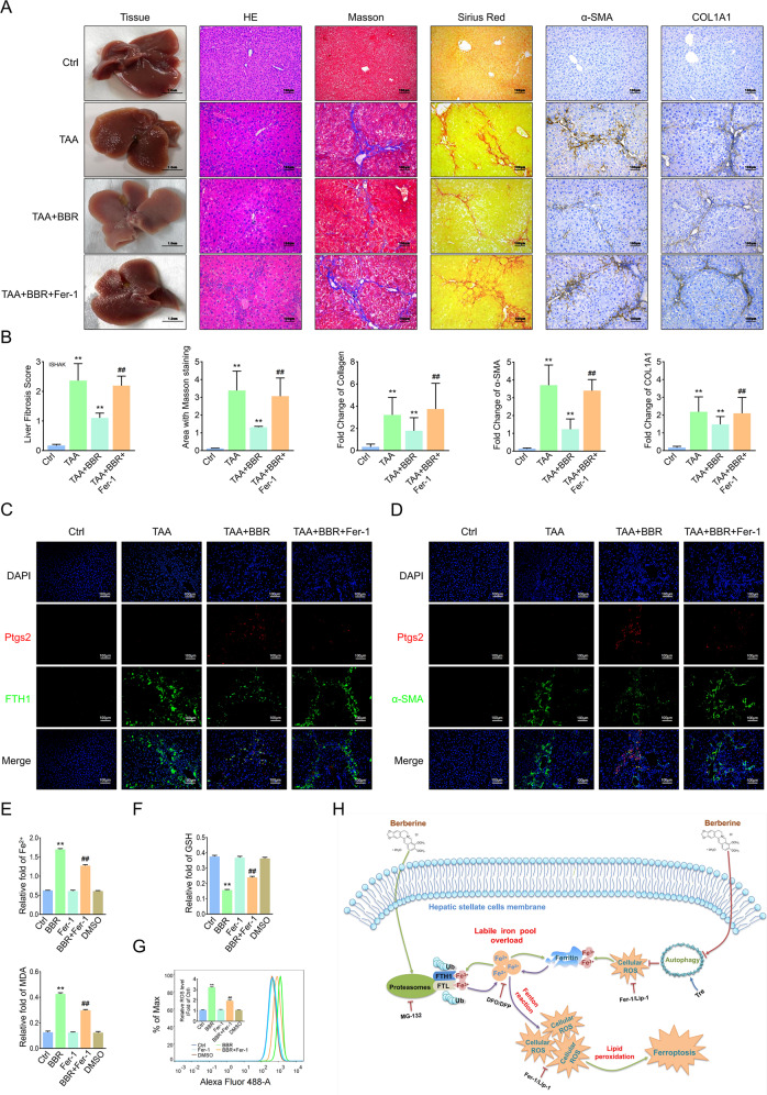
Fig. 8. Blockage of ferroptosis offsets the effect of BBR against liver fibrosis.
Mice were divided into 4 groups: control group, TAA-treated group, TAA-treated group plus BBR (200 mg/kg/day) with or without Fer-1 (1 mg/kg/day). A, B General examination of liver tissues by H&E, Masson’s trichrome, and Sirius red staining were performed. Fibrotic biomarkers COL1A1 and α-SMA staining in liver sections (compared with the control group, **P < 0.01; compared with the TAA plus BBR group, ##P < 0.01). C, D Mouse liver sections were costained with Ptgs2 and FTH1 or α-SMA. E–G HSC-LX2 was pretreated with Fer-1 (1 μM) for 1 h; then the cells were treated with BBR (21 μM) for 24 h, and ferroptotic events were investigated. H The underlying mechanism of BBR-induced HSC ferroptosis in liver fibrosis. The results are expressed as mean ± SD. Data are representative of three independent experiments; n = 3–6 in every group; Scales: 100 μm. Representative photographs are shown. Compared with the control group, **P < 0.01; compared with the BBR group, ##P < 0.01.

