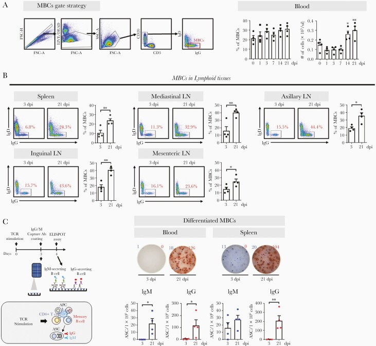
Figure 5.
MBCs in lymphoid tissues and blood. A, Representative gating strategy for MBCs. Frequency and absolute numbers of MBCs in the blood (1-way analysis of variance with Dunnett test for multiple comparisons vs 0 dpi; ∗P<.05, ∗∗P<.01). B, Frequency of MBCs in the spleen, mediastinal LN, axillary LN, inguinal LN, and mesenteric LN at 3 and 21 dpi (unpaired Student t test; ∗P<.05, ∗∗P<.01). Circles represent individual data obtained from each macaque and bars represent mean±SEM. C, Schematic diagram of the ELISPOT assay after TCR stimulation. SARS-CoV-2 spike protein (S1+S2)-specific IgM (blue) and IgG (red) ASCs were enumerated in MBCs activated by anti-CD3 and anti-CD28 monoclonal antibodies (1 μg/mL) in peripheral blood mononuclear cells (n=4) and the spleen (n=4) after 4-day culture. The ASCs were determined using the ELISPOT assay and are shown as mean±SEM (unpaired Student t test; ∗P<.05, ∗∗P<.01). Abbreviations: Ab, antibody; APC, antigen-presenting cell; ASC, antibody-secreting cell; dpi, days postinfection; ELISPOT, enzyme-linked immunospot; Ig, immunoglobulin; LN, lymph node; MBC, memory B cell; SARS-CoV-2, severe acute respiratory syndrome coronavirus 2; SEM, standard error of the mean; TCR, T-cell receptor.

