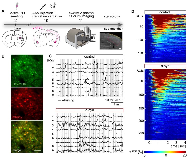
Figure 1.
Probing neuronal function in S1 of behaving mice upon striatal injection of a-syn PFFs. (A) Timeline of experiments. Mice received a striatal injection of PFFs at the age of 2 months, followed by the injection of AAV2/1.hSyn1.mRuby2.P2A.GCaMP6s into the somatosensory cortex (S1) and the implantation of a cranial window 8 months later when PFFs are globally present [assessed by immunofluorescence of phospho-synuclein (pSYN)]. One month later in vivo imaging experiments were conducted, after which mice were sacrificed and stereology was performed. (B) Representative example of a FOV. (C) Calcium traces of individual ROIs are shown for control and a-syn mice and referenced by whisking epochs (grey lines). (D) Heat maps depicting the average neural response to whisking onset (white line, normalized to the average activity within 0.5 s before whisking onset) for whisking responsive cells (control: 187 of 1561 neurons, a-syn: 276 of 1534 neurons). Scale bar in B 50 µm.

