Figure 4.
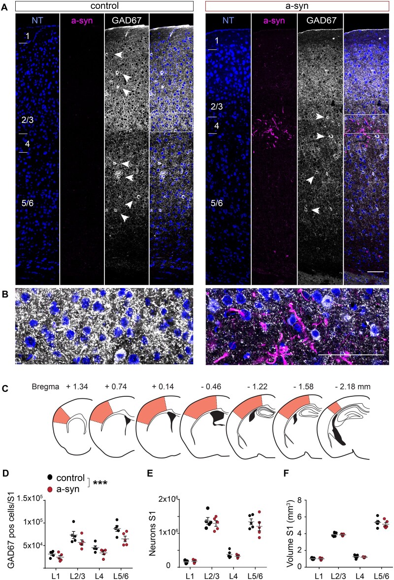
Stereology of somatosensory cortex reveals reduction of inhibitory neurons. (A) Representative examples of immunohistochemical stainings for neurotrace (NT, blue), phospho-synuclein (a-syn, magenta) and GAD67 (white arrow heads mark GAD67 positive GABAergic neurons) in S1 of a control and an a-syn PFF-seeded mouse assessed 9 months after striatal seeding. (B) Magnified area marked by a white box in (A). (C) Brain sections used for stereology to assess overall number of neurons and of GABAergic cells across all cortical layers in the entire S1 area (orange area). (D) The number of GAD67 positive interneurons was significantly reduced in a-syn mice [two-way ANOVA, effect of group F(1,32) = 14.69, P = 0.0006; effect of layer F(3,32) = 35.12, P < 0.0001; layer 5/6 P = 0.019; Bonferroni post hoc test, all other layers n.s], while neither (E) the total number of all neurons [effect of group F(1,32) = 1.41, P = 0.24, effect of layer F(3,32) = 132.2, P < 0.0001], nor (F) the cortical volume of S1 was affected [effect of group F(1,32) = 0.78, P = 0.39, effect of layer F(3,32) = 530.3, P < 0.0001]. Data are mean ± SEM in (D, E and F). Scale bar 100 µm in (A and B). *** P < 0.001.

