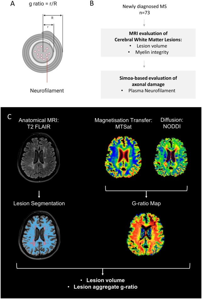
Figure 1.
Study overview: determining g-ratio of cerebral white matter lesions at multiple sclerosis diagnosis. Schematic cross-section of a myelinated axon, shown in A, showing definition of g-ratio, and location of neurofilaments. The g-ratio is defined as the ratio of the inner axonal radius to the outer fibre radius with the myelin sheath. Neurofilaments are critical structural proteins within the axon that are released into CSF and plasma in the context of axonal damage. Simultaneous evaluation of myelin integrity and axonal damage, shown in B, was performed in a cohort of 73 patients with newly diagnosed relapsing–remitting multiple sclerosis. A multimodal brain MRI protocol, shown in C, was acquired on a 3T clinical system (Prisma, Siemens, Erlangen.de), which included magnetization transfer saturation (MTsat) and multishell water diffusion MR acquisitions. WML were defined as hyperintensities on T2 Fluid-Attenuated Inversion Recovery and NAWM was segmented as described in the text. In this example, NAWM is shown in blue and WML in purple. Myelin integrity can be quantified as an ‘aggregate’ MR g-ratio, derived from MTsat and diffusion-derived neurite orientation dispersion and density imaging (NODDI) data, according to the equations shown in the text and Supplementary methods. The segmentation and g-ratio approaches were combined to determine aggregate g-ratios from both WML and NAWM.

