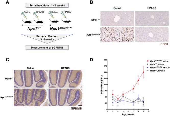
Figure 2 .

GPNMB levels are increased in Npc1m1N/m1N mutant mice and reduced by HPβCD treatment. (A) Experimental design. Mice received serial injections of HPβCD between 1 and 9 weeks of age, and serial plasma collection was performed for sGPNMB measurement by ELISA. (B) CD68 immunohistochemistry (brown staining) of liver sections demonstrated that the notable increase in foam cells characteristic of Npc1m1N/m1N mutant mice (lower left panel) is greatly reduced by HPβCD treatment (lower right panel). Saline-injected and HPβCD-treated Npc1+/+ controls are shown for comparison (upper left and right panels, respectively). Scale bar = 100 μm. (C) Immunostaining of midline sagittal sections of the cerebellum showed that elevated levels of GPNMB protein were present in 9-week-old saline-injected Npc1m1N/m1N mice (dark brown staining, lower left panel) when compared with the GPNMB levels in saline-injected Npc1+/+control mice (upper left panel). HPβCD-treated Npc1m1N/m1N mice (lower right panel) showed lower GPNMB expression in the cerebellum when compared with saline-injected Npc1m1N/m1N mice. HPβCD treatment of Npc1+/+ controls (upper right panel) did not affect GPNMB expression. Scale bar = 200 μm. (D) Levels of sGPNMB in plasma rose in untreated Npc1m1N/m1N mutant mice and were significantly different from Npc1+/+ controls. In contrast, sGPNMB levels remained low in HPβCD-treated mice and were not statistically different from those of Npc1+/+ mice. The mice shown in (D) were divided into two cohorts; see Supplementary Material, Fig. S6 for detailed, repeated measures data on individual mice, as well as a description of statistical analyses.
