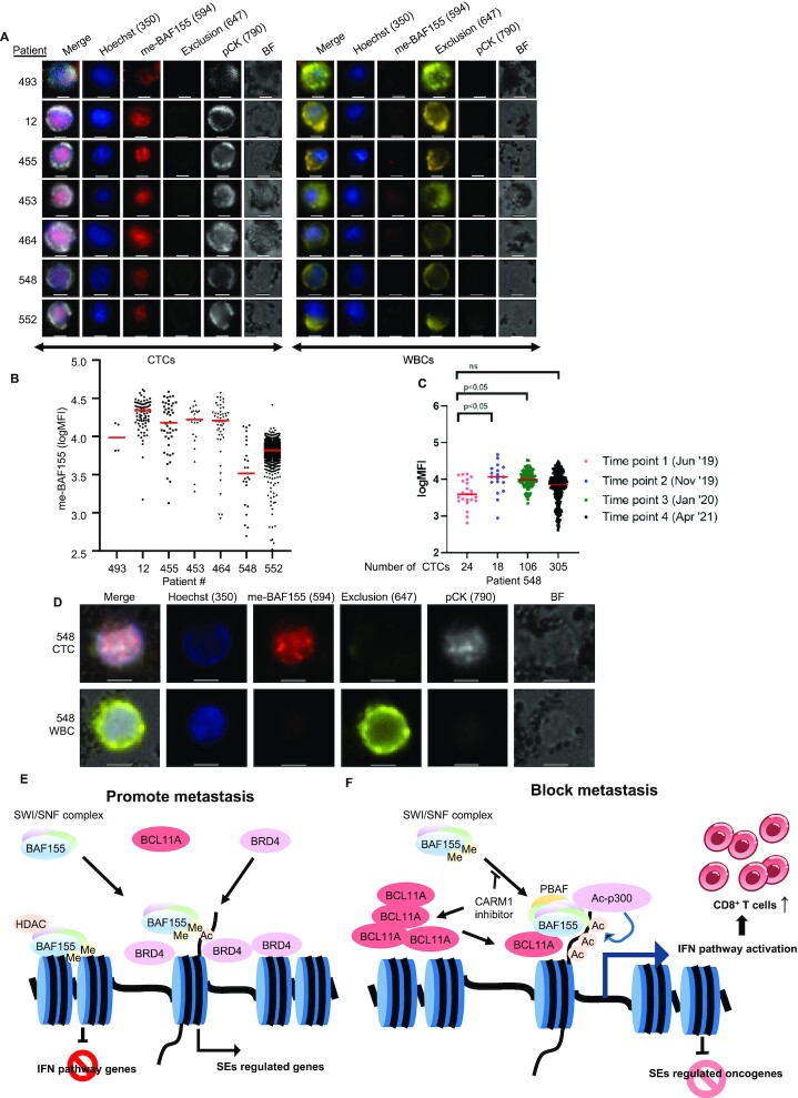
Figure 7.
Immunostaining of me-BAF155 in CTCs of metastatic breast cancer patients. (A) Images of me-BAF155 immunostaining in the nucleus of CTCs but not in WBCs from seven breast cancer patients. Representative images of CTCs (left) and WBCs (right) are shown for each patient (ID listed to the left). Each row of tiles includes an image crop of the intensity distribution of each of the stains included in the panel (Hoechst, me-BAF155, Exclusion (CD45/CD34/CD66b), pCK and Bright Field (BF)) for one single cell, including a merge of all stains. Images were taken at 20x magnification; scale bars represent 10 μm. (B) The average me-BAF155 nuclear signal intensities in breast cancer patient CTCs. Each dot represents the average me-BAF155 nuclear staining intensities of each individual CTC from one of the seven patients. The red bars indicate the average me-BAF155 nuclear signals of all CTCs from an individual patient sample. (C) The tracing of me-BAF155 nuclear signal intensities measurement in CTCs from breast cancer patient 548. The number of CTCs are depicted in x-axis. (D) Representative immunofluorescence images of CTCs stained with Hoechst, me-BAF155, Exclusion (CD45/CD34/CD66b), pCK and merged image of all staining signals for patient 548 in April 2021. Scale bars represent 10 μm. (E, F) Models depicting dual functions of me-BAF155 dependent metastasis in TNBC cells and anti-metastasis effects by CARM1 inhibition. Me-BAF155 and BRD4 interact at SEs to activate oncogenes and me-BAF155 containing SWI/SNF complex interacts with HDAC1 to suppress ISGs (E). CARM1 inhibitor treatment leads to increased BCL11A and un-methylated BAF155, which assemble with other SWI/SNF subunits to form PBAF to activate ISGs. Un-methylated BAF155 triggers dissociation of BRD4 from SEs, resulting in inhibition of expression of SE-addicted oncogenes (F).

