Figure 4.
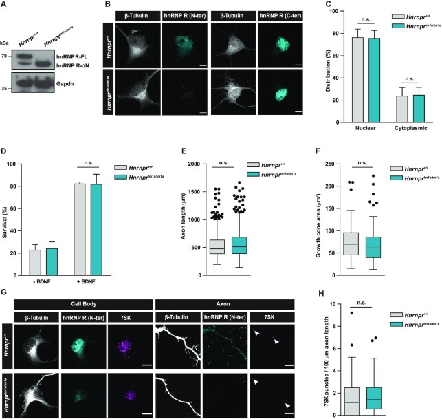
The hnRNP R-ΔN isoform is sufficient to support axon growth in primary motoneurons. (A) Western blot analysis of lysates from Hnrnpr+/+ and Hnrnprtm1a/tm1a motoneurons cultured for 7 DIV, probed with an antibody against the C-terminus of hnRNP R. Gapdh was used as loading control. (B) Representative images of primary motoneurons cultured for 7 DIV and immunostained with antibodies against the C-terminal or N-terminal domain of hnRNP R, and with an antibody against β-Tubulin. Scale bars: 5 μm. (C) Quantification of mean fluorescence intensity of the hnRNP R immunosignal obtained with the C-terminal-specific antibody in motoneurons shown in (B). Data are mean ± SD (n = 3 independent experiments; N = 30 motoneurons each for Hnrnpr+/+ and Hnrnprtm1a/tm1a). Statistical analysis was performed using two-way ANOVA followed by Bonferroni post-hoc test; n.s., not significant. (D) Quantification of the survival of motoneurons at 7 DIV as percentage of the number of motoneurons at 1 DIV, in the presence or absence of BDNF. Data are mean ± SD (n = 3 independent experiments; motoneurons were from N = 4 embryos each for Hnrnpr+/+ and Hnrnprtm1a/tm1a). Statistical analysis was performed using two-way ANOVA followed by Bonferroni post-hoc test; n.s., not significant. (E) Box-and-Whisker plots of the axon lengths of motoneurons cultured for 7 DIV. Data are mean ± SD (n = 3 independent experiments; N = 461 motoneurons for Hnrnpr+/+ and N = 502 motoneurons for Hnrnprtm1a/tm1a). Statistical analysis was performed using the Mann–Whitney test; n.s., not significant. (F) Box-and-Whisker plots of growth cone sizes of motoneurons cultured for 7 DIV. Data are mean ± SD (n = 3 independent experiments; N = 73 growth cones for Hnrnpr+/+ and N = 86 growth cones for Hnrnprtm1a/tm1a). Statistical analysis was performed using the Mann–Whitney test; n.s., not significant. (G) Representative images showing 7SK RNA labeling by in situ hybridization and immunostaining for hnRNP R in motoneurons derived from Hnrnpr+/+ and Hnrnprtm1a/tm1a mice. Arrowheads show 7SK-positive punctae. Scale bars: 5 μm. (H) Box-and-Whisker plots of the number of 7SK-positive punctae in axons of motoneurons cultured for 6 DIV. Data are mean ± SD (n = 3 independent experiments; N = 48 axons for Hnrnpr+/+ and N = 55 axons for Hnrnprtm1a/tm1a). Statistical analysis was performed using the Mann–Whitney test; n.s., not significant.

