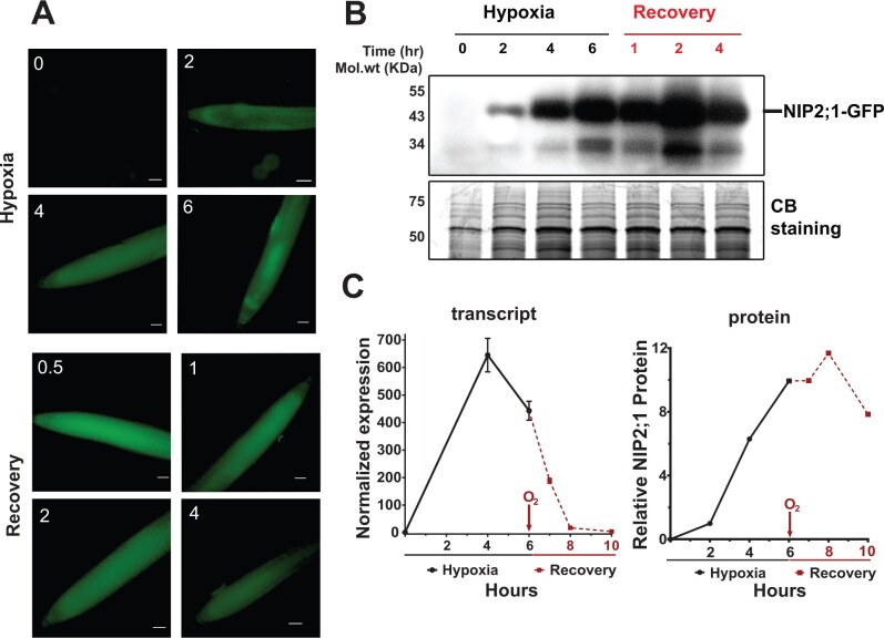
Figure 4.
NIP2;1-GFP expression in the roots of NIP2;1:GFP seedlings during hypoxia and reoxygenation. Ten-day-old NIP2;1:GFP seedlings were subjected to an argon-induced hypoxia time course, with oxygen resupplied at Hour 6. A, Representative epifluorescence images of the primary root of NIP2;1:GFP seedlings at the indicated times (h) of hypoxia treatment or reoxygenation recovery. Scale bars = 50 μm. B, Anti-GFP Western blot showing NIP2;1-GFP protein accumulation (upper). The expected position of NIP2;1-GFP is indicated. Bottom, Coomassie blue stained loading control gel. C, Comparison of root NIP2;1-GFP transcript levels (normalized to 0 h) by RT-qPCR (left, n = 8 biological experiments, error bars show sem) and NIP2;1-GFP protein (right) based on densitometry of the Western blot in (B).

