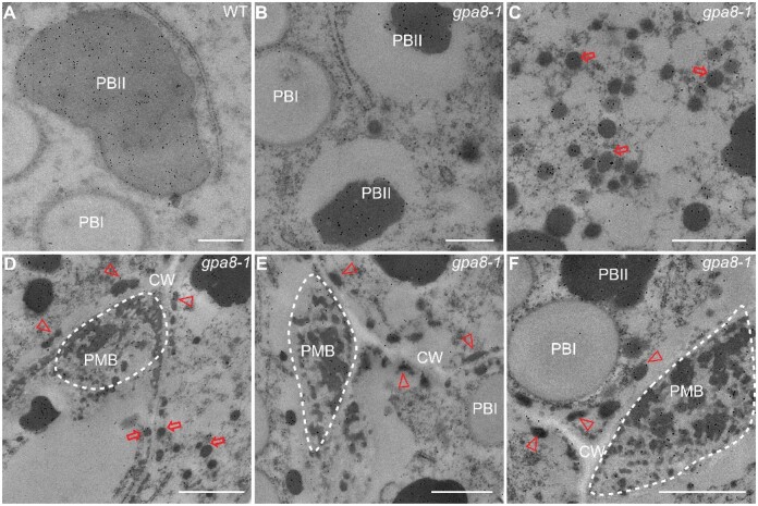
Figure 4.
Immunoelectron microscopy localization of glutelins in rice endosperm cells. A, Glutelins accumulated in PBIIs in WT endosperm cells. Bars = 500 nm. B, PBIIs in the gpa8-1 mutant were partially filled with glutelins. Bars = 500 nm. Irregular and round shape protein bodies indicate PBIIs and PBIs, respectively. C, Large clusters of DVs (red arrows) in the cytosol. Bars = 800 nm. D–F, Glutelins in DVs (red arrows), electron-dense granules (arrowheads), and PMB structures (dotted boxes) in gpa8-1. CW, cell walls. Bars = 1 μm in (D) and (F), Bars = 800 nm in (E). The 10-nm gold particle conjugated secondary antibodies were used in (A–F).

