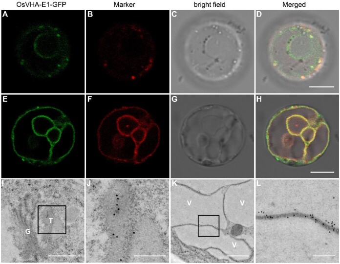
Figure 6.
Subcellular localization of OsVHA-E1. A–H, Subcellular localization of OsVHA-E1 in protoplasts of gpa8-1-complemented plants. OsVHA-E1-GFP were localized to the TGN and tonoplast. I–L, Subcellular localization of OsVHA-E1 in root tip cells of gpa8-1-complemented plants. Sections were prepared by the HPF/FS method. Immunoelectron microscopy analysis with anti-GFP antibodies revealed that OsVHA-E1-GFP were also localized to the TGN and tonoplast. Bars = 8 μm in (A–H). Bars = 600 nm in (I), 200 nm in (J, L), and 800 nm in (K). G, Golgi, T, TGN, V, vacuole. The 10-nm gold particle conjugated secondary antibodies were used in (I–L).

