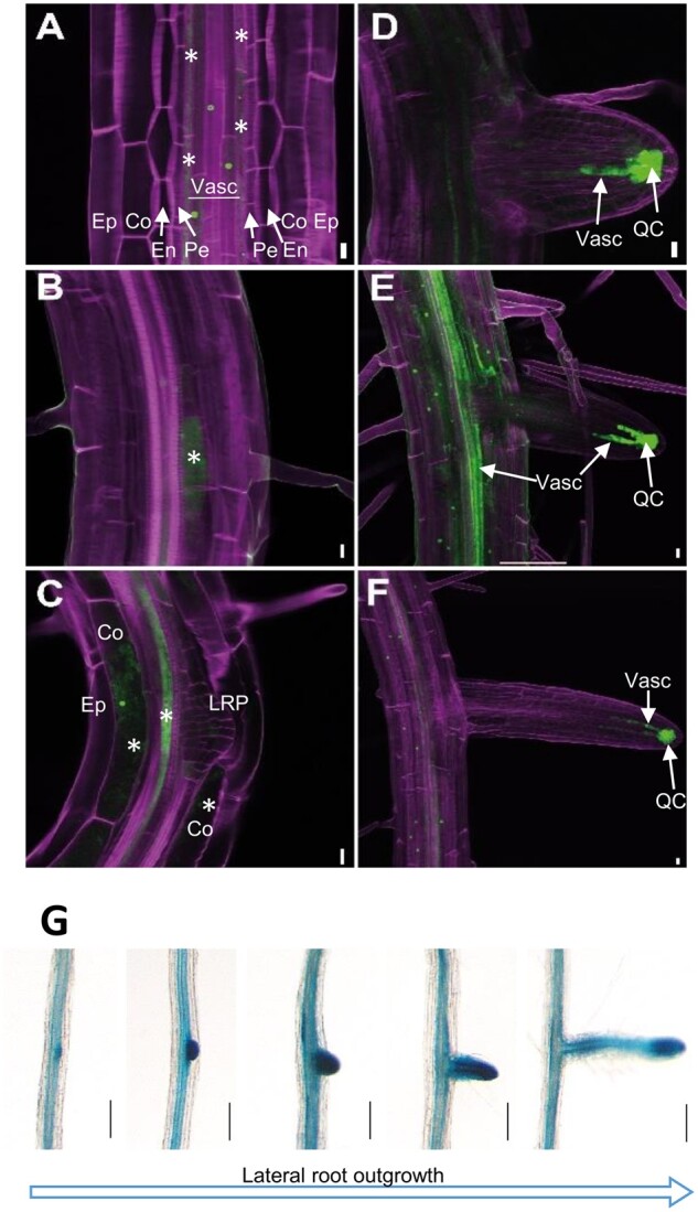
Figure 3.

ProSWEET17: HF-RPL18-GFP seedlings were grown for 7 d in 1/2 MS medium. A–F, Confocal images of different stages of LR development. In (A–C), pictures are single confocal optical sections plane and in (D–F), pictures are maximum intensity images from stacks of confocal optical sections. The calcofluor and GFP fluorescence are color-coded in magenta and green respectively. Co, cortex; Ep, epidermis; En, endodermis; QC, quiescent center and surrounding cells; Pe, pericycle; Vasc, vascular system. Scale bars represent 10 µm. Asterisks marks GFP fluorescence in cells adjacent to the pericycle layer (A), in cells in close vicinity to LRP (B and C), and in the cortex (C, Co). G, Histochemical localization of ProSWEET17-GUS activity during LR formation at different developmental stages detected from a root of single plant. ProSWEET17-GUS seedlings were grown for 14 d in MS medium followed by staining for GUS activity. Bars are 40 µm.
